

Sex differences identified in deadly brain tumors


For decades, scientists have recognized that more males get cancer and die of the disease than females. This is true for many types of cancer, including the deadly brain tumor glioblastoma. Now, a team of researchers led by Washington University School of Medicine in St. Louis has identified distinct molecular signatures of glioblastoma in men and women that help explain such underlying disparities in patients’ response to treatment and survival.

The research suggests that tailoring treatments to men and women with glioblastoma based on the molecular subtypes of their tumors may improve survival for all patients.

The findings are published Jan. 2 in Science Translational Medicine.

“It is our expectation that this study could have an immediate impact on the care of patients with glioblastoma and further research, as the findings indicate we should be stratifying male and female glioblastoma into risk groups and evaluating the effectiveness of treatment in a sex-specific manner,” said Joshua B. Rubin, MD, PhD, a Washington University professor of pediatrics and of neuroscience and the study’s co-senior author. “The biology of sex differences and its applications in medicine are highly relevant but almost always ignored aspects of personalized treatments.”

Glioblastoma is the most common malignant brain tumor and kills about half of patients within 14 months of diagnosis. It is diagnosed nearly twice as often in males, compared with females.

The tumor is most often diagnosed in people over age 50, and standard treatment is aggressive — surgery, followed by chemotherapy and radiation. However, stubborn stem cells often survive and continue to divide, producing new tumor cells to replace the ones killed by treatment. Most tumors recur within six months.

Studying adults with glioblastoma, the researchers found that standard treatment for glioblastoma is more effective in women than men.

To help understand such sex differences in treatment response, the researchers, including Kristin R. Swanson, PhD, a mathematical oncologist at the Mayo Clinic, measured tumor growth velocity in standard MRI scans.

“Basically, you can look at tumor growth velocity while patients are undergoing treatment and derive a value for how fast their tumors are growing,” said Rubin, who also is co-founder and co-director of the Pediatric Neuro-Oncology Program at St. Louis Children’s Hospital, where he treats patients. “This gives you an opportunity to think more deeply about whether the drug you’re giving a patient is actually helping.”

The researchers culled patient MRI scans and survival data from a cancer research database. They then calculated tumor growth velocity every two months for the duration of therapy in 63 glioblastoma patients — 40 males and 23 females — who received standard chemo-radiation treatment following surgery.

While initial tumor growth velocities were similar between females and males, only the females showed a steady and significant decline in tumor growth after treatment with temozolomide, the most common chemotherapy drug used to treat glioblastoma.

“The males did not respond as well, and we wanted to understand why, so we looked at the underlying genetics of patients’ tumors,” said Rubin, a co-leader of the Solid Tumor Therapeutics Program at Siteman Cancer Center at Barnes-Jewish Hospital and Washington University School of Medicine.

The researchers tapped into The Cancer Genome Atlas (TCGA) — a project launched in 2005 to pursue the genetic basis of cancer and funded by the National Cancer Institute and National Human Genome Research Institute, both of the National Institutes of Health (NIH). Led by the study’s co-senior author Jingqin “Rosy” Luo, PhD, a Washington University associate professor of surgery in the Division of Public Health Sciences, and the study’s lead author, Wei “Will” Yang, PhD, a Washington University bioinformaticist in the Department of Genetics, the researchers applied sophisticated statistical algorithms to distinguish  male- or female-specific gene expression patterns from such patterns that were shared among the male and female patients. The team then focused on the sex-specific gene expression to identify molecular subtypes that corresponded to differences in survival for males and for females.

“We observed tremendous genetic sex differences in the tumors of glioblastoma patients that correlated with survival,” Luo said. “All evidence supports the need to define these distinctions and incorporate the sex differences into glioblastoma biology research and treatment.”

Specifically, the researchers showed that the tumors of patients with glioblastoma cluster into 10 distinct subtypes — five for tumors in males and five for tumors in females. The clusters are distinguished by gene activity and survival. For example, females with tumors in one such cluster survived longer than females with tumors in any of the other four clusters — just over three years compared with just over one year. Similarly, they found a male cluster linked to longer survival — just over 18 months compared with just over one year for men with tumors in the other clusters.

The researchers validated the clusters in three additional data sets and also showed that even genes activated at similar levels in tumors in males and females can result in substantial sex-specific effects on survival.

“Additionally, we identified genetic pathways that correlated with the longest survival, and they were very different in males compared with females,” Rubin said. “For example, in males survival was all about regulating cell division, which suggests that drugs that block cell-cycle progression may be more effective in men. For females, survival was all about regulating invasiveness, which suggests that drugs targeting integrin signaling may be more effective in women. This tells us it might be better to separate males and females and examine their sex-specific genetic signatures. We tested this hypothesis by doing a series of in vitro drug screens in which we took four relatively common chemo drugs and looked at how the expression of these genes correlated with response to those drugs. In both males and females, there was a clear correlation.”

Among diseases in general, sex differences are often tied to hormones. For example, the female hormone estrogen contributes significantly to more women getting breast cancer than men. However, with glioblastoma diagnosis and survival, sex hormones did not directly contribute to female and male differences, Rubin said. “The sex-specific genetic activity in glioblastoma is not dependent on the acute actions of circulating sex hormones as differences are evident across all stages of life.”

“In a broader sense, I want our research to encourage people to think more about how diseases uniquely affect males and females, making it the norm and not the exception,” Rubin added. “I hope the research will inspire more specific approaches to treatments. It may be that we shouldn’t be using the same criteria when treating diseases in males and females, and as a next step we should definitely develop and evaluate sex-specific treatment regimens for glioblastoma.”

In addition to researchers at Washington University and the Mayo Clinic, scientists at the Cleveland Clinic, Case Western Reserve University and TGen, a genomics research institute, also contributed to the study.
$5 million supports innovative breast cancer trial at Siteman

A $5 million grant from the Department of Defense will support research at Washington University School of Medicine in St. Louis aimed at improving breast cancer therapies. The study, which includes a clinical trial at Siteman Cancer Center at Barnes-Jewish Hospital and the School of Medicine, will focus on HER2-positive breast cancer. Such tumors are dotted with an overabundance of so-called HER2 receptors.

About 20 percent of women with breast cancer have HER2-positive tumors. Drugs that block HER2, such as Herceptin, have improved survival rates dramatically for these patients.

But these drugs’ use has been limited to patients who have too many copies of HER2. Recent studies led by Washington University researchers, however, have shown that other breast cancer patients with different HER2 defects may benefit from HER2 inhibitors. In these patients, mutations in HER2 can fuel cancer growth. Standard testing for HER2 positive breast cancer won’t identify patients with HER2 mutations.

“We’re figuring out how to treat breast cancer based on the mutations present in the specific patient’s tumor,” said co-principal investigator Ron Bose, MD, PhD, an associate professor of medicine and Siteman research member. “We’ve developed a diagnostic test for mutations in HER2 that cause overactive signaling, and we have a good drug for patients with these mutations in their tumors. These new diagnostic and therapeutic tools may let us identify more women with HER2-driven cancers and treat them with HER2 inhibitors.”

Based on data from 2017, Bose and his colleagues estimate that about 4,000 women have metastatic breast cancer with HER2 mutations and could potentially benefit from this strategy. Drug combinations targeting tumor proteins are generally less toxic than standard chemotherapy, which is not very effective in controlling metastatic cancer and is the only current treatment option for these patients.

The clinical trial at Siteman will investigate a HER2 blocker called neratinib. Trial participants will be patients with metastatic breast tumors that have HER2 mutations and are estrogen-receptor positive, meaning these tumors also are fueled by estrogen. Because these tumors are fed by two fuel sources, participants will receive neratinib to block HER2 and fulvestrant to attack the estrogen receptor.

“In a past clinical trial, we tested neratinib alone, and we saw that about 30 percent of patients had a positive therapeutic benefit from the drug,” said co-principle investigator Cynthia X. Ma, MD, PhD, an associate professor of medicine and Siteman research member. “Now, we want to see whether adding fulvestrant will improve that outcome and help more patients.”

To more fully understand how patients respond to the drugs, the researchers will implant the patients’ tumors into mice that then will receive the same treatment regimens. Additional drugs also will be tested in these mouse models to see if other treatment combinations suggest promising directions for future clinical trials.

The mouse models will provide insight into why some tumors become resistant to neratinib therapy. The scientists will perform genome sequencing and protein analysis on these tumors, seeking clues to how some of them develop the ability to evade drugs that had been lethal to them initially.

Collaborating sites for the clinical trial include Dana-Farber Cancer Institute at Harvard Medical School and Baylor College of Medicine.

This work is supported by the U.S. Department of Defense, grant numbers 12473122 and 12473075.

Brain cancer vaccine effective in some patients

Most people with the deadly brain cancer glioblastoma die less than 18 months after diagnosis. But a multicenter clinical trial of a personalized vaccine that targets the aggressive cancer has indicated improved survival rates for such patients.

The study appears May 29 in the Journal of Translational Medicine.

The phase three clinical trial included 331 patients at over 80 sites in four countries. Among the trial participants was Siteman Cancer Center at Barnes-Jewish Hospital and Washington University School of Medicine in St. Louis, which recruited one of the largest groups of patients in the trial. The patients were randomized to receive standard therapy plus the personalized vaccine, called DCVax-L, or standard therapy plus a placebo. Personalized vaccines are specifically tailored to individual patients.

According to the trial design, the vaccine group had twice the number of patients as the placebo group. Further, any patient in the trial was allowed to receive the vaccine if his or her cancer recurred or progressed after initial treatment. As such, almost 90 percent of all participants received the investigational vaccine.

The trial, which was funded by the vaccine maker, Northwest Biotherapeutics Inc., is ongoing to allow for continued study of patients who are living beyond what is expected of those diagnosed with glioblastoma.

As of the analysis detailed in the study, all 331 patients in the trial (including those who did and did not receive the vaccine) had a median survival of just over 23 months. One-hundred patients had an average overall survival of 40.5 months — more than three years — and were designated as “extended survivors.” Investigators are particularly interested in the latter group’s response to the vaccine, though they don’t yet know whether everyone in this group received the vaccine since the trial is ongoing and remains blinded. The continued blinding means neither the patients nor physicians know which patients are receiving the vaccine. But since the researchers reported that almost 90 percent of all participants received the vaccine at some point, the chances that the extended survivors are getting the vaccine, rather than placebo, are high.

“The overall patient population in the trial appears to live longer than we would typically see with current standard of care, and 30 percent of the patients have lived much longer than we would expect, given the typical course of this cancer,” said oncologist Jian L. Campian, MD, PhD, one of the study’s authors and a Washington University assistant professor of medicine who treats patients at Siteman Cancer Center. “In general, patients with this cancer live 15 to 17 months. The surprising part was that the 100 ‘extended survivors’ don’t appear to have the usual characteristics associated with a good prognosis. We are continuing to study these patients to understand why they have done so well.”

The personalized vaccine used was specific to each patient. After surgery to remove as much of the tumor as possible, a small amount of tumor tissue is processed and then exposed to the same patient’s own immune cells, called dendritic cells. Exposure to the tumor material essentially trains the dendritic cells to seek out and destroy tumor cells. These trained dendritic cells are returned to the patient as a vaccine injected in the arm. Relatively few vaccinations are needed: The first three are weeks apart; the next three are months apart; and then they continue with one vaccination every six months after the first year.

Campian said the vaccine had very few side effects, especially compared with standard treatment for this cancer, which includes surgery, radiation and chemotherapy. About 2 percent of participants (seven patients) experienced a serious adverse event — such as brain swelling or seizures — that may have been related to the vaccine, according to the researchers.

Experimental arthritis drug prevents stem cell transplant complication

An investigational drug in clinical trials for rheumatoid arthritis prevents a common, life-threatening side effect of stem cell transplants, new research from Washington University School of Medicine in St. Louis shows. Studying mice, the researchers found the drug prevented what’s known as graft-versus-host disease, a debilitating, sometimes lethal condition that develops when transplanted stem cells attack the body’s own organs or tissues.

About half of patients receiving donor stem cells develop graft-versus-host disease, which can linger for months or years after their transplants. In some cases, patients die not from their cancer but from the complication itself. Current treatments are not effective.

The study is online in the journal Leukemia.

In past work, this research team defined the role of molecules called JAK1/2 kinases and their signaling pathways in immune cell activation and graft-vs-host disease. In the new study, these same researchers evaluated ruxolitinib and baricitinib, and found baricitinib to be the superior of the two drugs in reducing and preventing graft-versus-host-disease in mice. Both drugs belong to a class of pharmaceuticals called JAK inhibitors that are known for dialing down inflammation.

“Transplanted donor stem cells — and more specifically, the T cells in the donor stem cell product — are particularly good at fighting off leukemia, but these cells can go haywire, unfortunately, and attack the patient’s healthy tissues, causing graft-versus-host disease,” said senior author John F. DiPersio, MD, PhD, the Virginia E. and Sam J. Golman Professor of Medicine in Oncology. “The typical ways we can reduce the effects of the disease also tend to weaken the T cells’ ability to attack the cancer. We’re looking for a treatment strategy that stops the disease without shutting down T cells’ assault on the cancer.”

Surprisingly, baricitinib did more than shut down graft-versus-host disease. It actually boosted the ability of the donor T cells to fight the cancer.

“We don’t know yet exactly how this happens, but we’re working to understand it,” said first author Jaebok Choi, PhD, an assistant professor of medicine. “We think at least part of the explanation is the drug strips the leukemia cells of their immune defenses, making them more vulnerable to attack by the donor T cells. At the same time, the drug also stops the donor T cells from being able to make their way to important healthy tissues, such as the skin, liver and gastrointestinal tract, where they often do the most damage.”

In other words, the drug appears to stop graft-versus-host disease by simply keeping the donor T cells circulating in the bloodstream, away from vital organs. Simultaneously, the drug makes the leukemia cells more vulnerable to immune attack from the donor T cells, which are now mostly confined to the bloodstream, where the cancer is.

The drug also appeared to boost levels of specific immune cells that put the brakes on a runaway immune response that can make graft-versus-host disease worse. These apparently independent effects are specific to baricitinib and may explain why other JAK inhibitors did not work as well, according to DiPersio, who is also deputy director of Siteman Cancer Center at Barnes-Jewish Hospital and Washington University School of Medicine.

The researchers emphasized the finding that the drug not only prevented graft-versus-host disease from developing in the mice but reversed established disease, suggesting possible options for patients already affected by it.

“We were surprised to achieve 100 percent survival of mice with the most severe model of graft-versus-host disease,” Choi said. “We are now studying the multi-pronged ways this drug behaves in an effort to develop an even better version for eventual use in clinical trials.”

Because of the drug’s broad effectiveness in preventing inflammatory disorders, DiPersio said he and his colleagues are beginning to explore whether it could be used to prevent organ rejection in patients undergoing solid organ transplantation. Such a strategy might reduce the need to give these patients powerful immune-suppressing drugs that increase the risk of infection.

For women with kidney cancer, belly fat matters

Belly fat affects the odds of women surviving kidney cancer but not men, according to a new study by researchers at Washington University School of Medicine in St. Louis.

Half of female kidney cancer patients with substantial abdominal fat at the time of diagnosis died within 3 1/2 years, while more than half of women with little belly fat were still alive 10 years later, the researchers found. For men, the amount of abdominal fat appeared to make no difference in how long they survived.

The findings suggest that kidney cancer may develop and progress differently in women than men.

“We’re just beginning to study sex as an important variable in cancer,” said senior author Joseph Ippolito, MD, PhD, an instructor in radiology at Mallinckrodt Institute of Radiology at the School of Medicine. “Men and women have very different metabolisms. A tumor growing in a man’s body is in a different environment than one growing inside a woman, so it’s not surprising that the cancers behave differently between the sexes.”

The study is available online in the journal Radiology.

Excess weight is a major risk factor for the development of kidney cancer, but it does not necessarily portend a poor outcome. Rather, the new study suggests that how long a patient survives after diagnosis is linked not to total fat but to the distribution of body fat, at least for women.

Most methods of estimating body fat rely on just a person’s height and weight. But not all fat is the same. The kind you can squeeze – called subcutaneous fat – seems to be mostly harmless. But visceral fat, which lies within the abdomen and encases internal organs, has been associated with diabetes, heart disease and many kinds of cancer.

Visceral fat sits too deep inside the abdomen to be measured accurately with a tape measure around a person’s waist. Instead, Ippolito and colleagues analyzed cross-sectional CT scans, which are routinely performed on people newly diagnosed with kidney cancer to measure the size of tumors and to look for metastases. Subcutaneous and visceral fat are located in different areas of the body on a CT scan, making it possible to calculate the proportion of each.

The researchers analyzed images from 145 men and 77 women with kidney cancer. The scans were drawn from The Cancer Imaging Archive, a collection of demographic, clinical and imaging data on hundreds of cancer patients.

The researchers found that half of the women with high visceral fat died within 3 ½ years of diagnosis, while more than half of the women with low visceral fat were still alive after 12 years. Women often gain visceral fat after menopause, but the link still held after correcting for age.

For men, there was no correlation between visceral fat and length of survival.

“We know there are differences in healthy male versus healthy female metabolism,” Ippolito said. “Not only in regard to how the fat is carried, but how their cells use glucose, fatty acids and other nutrients. So the fact that visceral fat matters for women but not men suggests that something else is going on besides just excess weight.”

That “something else” could lie in the tumor cells themselves. Tumor cells prefer sugar as a fuel source, but some have more of a sweet tooth than others. A sugar-hungry tumor typically spells trouble for patients.

Using data from The Cancer Genome Atlas, the researchers analyzed the gene expression profiles of tumors from 345 men and 189 women diagnosed with kidney cancer. Both men and women were less likely to survive if their tumor cells had switched on the genes associated with consuming sugar, or glycolysis. Men whose tumor cells exhibited low glycolysis survived an average of 9 ½ years, whereas those with high-glycolysis tumors survived for only six years on average.

The researchers found 77 women with matched imaging and gene expression data, so they combined their analyses of visceral fat and glycolysis.

About a quarter of the women had a high amount of visceral fat and tumors whose glycolysis genes were significantly active. Those women survived only two years after diagnosis on average. Strikingly, of the 19 women who fell into the low visceral fat and low glycolysis category, none died before the end of the study, which covered a span of 12 years. There was no group of men with a similarly rosy prognosis.

“We found there’s a group of women that’s doing really poorly relative to everyone else, and a group that’s doing really well,” Ippolito said. “Our data suggest that there is a potential synergy between the patient’s visceral fat and the metabolism of their tumor. That can be a starting point to figure out how to better treat women with kidney cancer. We would not have discovered this if we had been looking at men and women together.”

Major milestone reached in effort to ID cancers’ genetic roots

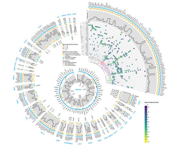
Researchers nationwide have reached a major milestone in describing the genetic landscape of cancer. Scientists at Washington University School of Medicine in St. Louis and other institutions have completed the genetic sequencing and analyses of more than 11,000 tumors from patients, spanning 33 types of cancer — all part of The Cancer Genome Atlas (TCGA) project, funded by the National Cancer Institute and National Human Genome Research Institute, both of the National Institutes of Health (NIH).

Altogether, the researchers identified about 300 genes that drive tumor growth. And, remarkably, just over half of all tumors analyzed carry genetic mutations that could be targeted by therapies already approved for use in patients.

The Circos plot shows the 299 genes identified as drivers of cancer and summarizes many other pieces of data from TCGA. Briefly, the outer text in blue indicates different cancer types. The predicted driver genes unique to that cancer type are listed in black text. The top right section shows all genes found to be important in multiple cancer types.


TCGA was launched in 2005 to pursue the genetic basis of cancer. The results appear April 5 in a series of studies published in Cell Press journals.

“For the 10,000 tumors we analyzed, we now know — in detail — the inherited mutations driving cancer and the genetic errors that accumulate as people age, increasing the risk of cancer,” said leading TCGA scientist Li Ding, PhD, an associate professor of medicine and director of computational biology in the Division of Oncology at Washington University. “This is the first definitive summary of the genetics behind 33 major types of cancer.”

Ding, also an assistant director of  The McDonnell Genome Institute at Washington University, is a major author of six papers published April 5 in the journals Cell, Cell Reports and Cell Systems, detailing the genetic mutations underlying cancer.

Genomic studies over the past decade have demonstrated that cancer is a disease of errors in genes rather than particular organs.

“This project is the culmination of more than a decade of groundbreaking work,” said NIH Director Francis S. Collins, MD, PhD. “This analysis provides cancer researchers with unprecedented understanding of how, where, and why tumors arise in humans, enabling better-informed clinical trials and future treatments.”

Ding said the new analyses have revealed that the genetic errors of cancer result in specific molecular signatures that could guide treatment.

“Rather than the organ of origin, we can now use molecular features to identify the cancer’s cell of origin,” Ding said. “We are looking at what genes are turned on in the tumor, and that brings us to a particular cell type. For example, squamous cell cancers can arise in the lung, bladder, cervix, and some tumors of the head and neck. We traditionally have treated cancers in these areas as completely different diseases. But studying their molecular features, we now know such cancers are closely related. Cancers originating in, for example, epithelial cells that line various organs are similarly closely related, regardless of their location.”

According to Ding, the research supports the idea that tumors of any type with high numbers of mutations — which often are resistant to chemotherapy — are susceptible to immunotherapy drugs called checkpoint inhibitors. Highly mutated tumors produce comparatively more misshapen proteins that can trigger an immune response. But as a safeguard against autoimmunity, the body often puts the breaks on such an immune response. Still, to treat aggressive tumors, checkpoint inhibitors can remove those breaks, letting the immune system fight the tumor more effectively.

The studies also provide further clarity on the significance of certain mutations in the BRCA1 gene that drive breast and ovarian cancer. Specific mutations in this gene are known to significantly increase the risk of certain types of cancer. But the consequences of many other mutations in this gene were unknown, making it difficult to predict cancer risk.

“We have known for a long time that BRCA1 is an important gene in cancer development,” said Ding, who has a research affiliation with Siteman Cancer Center at Barnes-Jewish Hospital and Washington University School of Medicine. “But it’s very hard to tease out which specific mutations in BRCA1 are actually driving the cancer and which mutations are harmless. Our paper on cancer-causing variants in inherited mutations provides new clarity on the BRCA1 mutations actually driving tumor growth. We found 21 disease-causing BRCA1 and BRCA2 variants in breast cancer, three in cervical cancer, one in colorectal cancer, one in glioblastoma and 38 in ovarian cancer.”

Ding said she is particularly excited about the prospect of using these analyses to re-examine data from past clinical trials. Many times, a small proportion of patients in a given trial did well on an experimental therapy, but many others did not respond to the treatment at all, and researchers didn’t understand why. Perhaps a drug was not approved for, say, lung cancer because of such results, but some patients with certain cancer mutations may benefit.

“Most earlier trials were not designed with genomics in mind,” Ding said. “We know how these patients responded. Now, we can sequence the tumor samples from patients enrolled in those trials with our latest software tools. We can look for correlations between the patients’ genomics and how they responded to the treatments. If we do this for many past trials, we will have tremendous statistical power to identify reasons why drugs work for some patients and not others. So even negative trials that might have been a disappointment at the time can become powerful tools to design better treatments in the future.”

In this way, a drug that might have failed as a treatment for lung cancer might be re-examined as a potential therapy for, say, squamous cell carcinoma, again, regardless of location.

“Even after genomic sequencing, sometimes we still can’t explain what is going on,” Ding said. “This is why we are planning to expand beyond studies of the tumor cells to include the entire tumor ecosystem — the immune cells that infiltrate the tumor and the supporting tissue that creates the tumor’s microenvironment.”

Added Eric Green, MD, PhD, director of the National Human Genome Research Institute, “These TCGA publications effectively showcase how genomics has brought transformative advances to the study of cancer. Central to these advances has been the development and use of new and increasingly more powerful technologies and approaches for genome sequencing.”

TCGA is a collaborative effort involving more than 20 institutions. The McDonnell Genome Institute and many researchers at Washington University have made major contributions to this national project. In the latest batch of seminal papers, Kuan-lin Huang, Matthew H. Bailey, Reyka G. Jayasinghe, Qingsong Gao, Song Cao, Wen-Wei Liang, Steven M. Foltz and others from Ding’s lab are primary authors. Within Washington University, Ding has collaborated with a number of investigators, including Feng Chen, PhD, an associate professor of medicine and a co-senior author of two of these papers.

CRISPR enhances cancer immunotherapy

Last year, the Food and Drug Administration approved the first cellular immunotherapies to treat cancer. These therapies involve collecting a patient’s own immune cells — called T cells — and supercharging them to home in on and attack specific blood cancers, such as hard-to-treat acute lymphoblastic leukemia and non-Hodgkin lymphoma.

But so far, these T cell immunotherapies — called CAR-T cells — can’t be used if the T cells themselves are cancerous. Even though supercharged T cells can kill cancerous T cells, they also can kill each other because they resemble one another so closely.

Scientists at Washington University School of Medicine in St. Louis now have used the gene-editing technology CRISPR to engineer human T cells that can attack human T cell cancers without succumbing to friendly fire.

The study evaluating the approach in mice appears online in the journal Leukemia.

The researchers also engineered the T cells so any donor’s T cells could be used. A “matched” donor with similar immunity is not required and neither are the patient’s own T cells, which is important for the obvious reason: Many of the patient’s own T cells are cancerous.

“Cancerous T cells and healthy T cells have exactly the same protein — CD7 — on their surfaces,” said senior author John F. DiPersio, MD, PhD, the Virginia E. and Sam J. Golman Professor of Medicine in Oncology.

DiPersio’s team first generated a novel CAR-T strategy targeting CD7, allowing for the targeting and killing of all cells with CD7 on the surface.

“But if we program T cells to target CD7, they would attack the cancerous cells and each other, thus undermining this approach,” DiPersio said. “To prevent this T cell fratricide, we used CRISPR/Cas9 gene editing to remove CD7 from healthy T cells, so they no longer carry the target.”

DiPersio, who treats patients at Siteman Cancer Center at Washington University School of Medicine and Barnes-Jewish Hospital, and his colleagues also used CRISPR gene editing to simultaneously eliminate the therapeutic T cells’ ability to see healthy tissues as foreign.

To do this, they genetically deleted the T cell receptor alpha (TCRa) subunit. This way, T cells from any normal donor can be used without risk of life-threatening toxicities such as graft-versus-host disease, in which T cells attack the organs of the recipient, sometimes resulting in death. This new approach also may have broad implications for the CAR-T field, allowing for use of therapeutic T cells from any healthy donor. Healthy T cells could be collected in advance and stored for any patient with a relapsed T cell malignancy.

“We have genetically modified these T cells so they are unable to cause graft-versus-host disease but can still kill cancerous cells,” said first author Matthew L. Cooper, PhD, an instructor in medicine. “One additional benefit of this approach is that a patient could receive this therapy much more quickly after diagnosis. We wouldn’t need to harvest the patient’s own T cells and then modify them, which takes time. We also wouldn’t have to find a matched donor. We could collect T cells from any healthy donor and have the gene-edited T cells ready in advance, a strategy termed ‘off-the-shelf’ CAR-T cell therapy.”

The researchers demonstrated that this approach is effective in mice with T cell acute lymphoblastic leukemia (T-ALL) taken from patients. Mice treated with the gene-edited T cells targeted to CD7 survived 65 days, compared with 31 days in a comparison group that received engineered T cells targeting a different protein. The researchers also found no evidence of graft-versus host disease in mice that received T cells lacking the molecular machinery that sees healthy tissues as foreign. They also found that the therapeutic T cells remained in the blood for at least six weeks after the initial injection, suggesting it could ramp up again to kill cancerous T cells if they return.

“T cell malignancies represent a class of devastating blood cancers with high rates of relapse and death in children and adults with the disease,” Cooper said. “In an effort to develop the first clinically viable targeted therapy for this type of cancer, we are scaling up the manufacturing of our gene-edited CAR-T cells for clinical trials, which we hope to complete at Siteman Cancer Center.”

Higher doses of radiation don’t improve survival in prostate cancer

A new study shows that higher doses of radiation do not improve survival for many patients with prostate cancer, compared with the standard radiation treatment. The analysis, which included 104 radiation therapy oncology groups across North America, was led by researchers at Washington University School of Medicine in St. Louis.

Past studies have shown that gradually escalating the radiation dose resulted in improved cancer control, such as slower tumor growth and lower levels of prostate-specific antigen (PSA), an indicator of cancer growth. The new study, published March 15 in JAMA Oncology, is the first that is large enough to examine whether these improved measurements translate into longer survival for patients.

“Our goal is to improve survival, but we didn’t see that despite advances in modern radiotherapy,” said first author Jeff M. Michalski, MD, the Carlos A. Perez Distinguished Professor of Radiation Oncology. “But we did see significantly lower rates of recurrence, tumor growth and metastatic disease — tumors that spread — in the group that received the higher radiation dose. Still, that didn’t translate into better survival. The patients in the trial did better than we anticipated, and part of that may have been because of improvements in metastatic cancer therapy over the 10 years of the trial.”

The study included about 1,500 patients with intermediate-risk prostate cancer, the risk category in which most patients fall. To be classified in this risk category, patients generally have PSA scores of 10-20 ng/ml and a Gleason score of seven, the latter of which is a measure of tumor aggressiveness. Both treatment groups received external beam radiation. The standard group received a radiation dose of 70.2 gray delivered over 39 treatment visits. The investigational group received increasing doses up to 79.2 gray delivered over 44 visits. (A gray is the standard measure of radiation a material has absorbed.)

Of the 748 men receiving standard treatment, 75 percent were still alive after eight years of follow-up. Of the 751 men receiving the dose-escalation treatment, 76 percent were alive at the eight-year mark — a difference that is not statistically significant. These overall survival rates include deaths for any cause, not just those due to prostate cancer. Over the course of the study, 51 patients died of prostate cancer, which is 3.4 percent of all patients enrolled. At the eight-year mark, the death rate due to prostate cancer for patients receiving standard treatment was 4 percent compared with 2 percent for patients receiving the escalating dose. These rates also were not statistically different.

While there was no difference in overall survival numbers, Michalski pointed out some differences in side effects and in whether further treatment was needed later. Such differences could help doctors and patients in deciding the best treatment course.

Patients in the standard dose group were more likely to undergo further therapies to control tumors that had grown larger or that had spread to another site in the body. But patients in the escalating dose group experienced more side effects — such as urinary irritation or rectal bleeding — sometimes years after treatment.

During the 10 years it took to enroll enough patients in the trial, Michalski said, at least six new therapies were approved for recurrent or metastatic prostate cancer, and these therapies have been shown to improve survival. It is possible the patients in the standard treatment arm — who were shown to need more follow-up therapies — would not have done as well as the group receiving the escalating dose had these new therapies not become available.

“If there is a difference between standard and escalating doses, it’s hard to show it when the patients who later develop recurrent cancer can have their lives extended through the use of additional therapies,” said Michalski, who treats patients at Siteman Cancer Center at Washington University School of Medicine and Barnes-Jewish Hospital. “Of course, these additional therapies have their own side effects, as does the higher initial dose of radiation therapy. In addition, the selective use of androgen withdrawal therapy has been shown to improve survival in men treated with radiation therapy. This treatment can be combined with either standard or higher dose radiation therapy.”

“If we can safely deliver the higher dose of radiation, my opinion is to do that,” Michalski added. “It does show lower risk of recurrence, which results in better quality of life. But if we can’t achieve those ‘safe’ radiation dose goals, we shouldn’t put the patient at risk of serious side effects down the line by giving the higher dose. If we can’t spare the rectum or the bladder well enough, for example, we should probably back off the radiation dose. It’s important to develop treatment plans for each patient on a case-by-case basis.”

Cutting off cervical cancer’s fuel supply stymies tumor

Cancer therapies have improved — in some cases dramatically — over the past two decades, but treatment for cervical cancer has remained largely unchanged. All patients receive radiation and chemotherapy, yet despite the aggressive approach, the regimen fails in about one-third of patients with cervical cancer that has spread beyond the cervix but not outside the pelvis.

Now, researchers at Washington University School of Medicine in St. Louis have shown that cervical tumors that don’t respond to radiation may be vulnerable to therapies that also attack the cancer’s fuel supply. Studying mice implanted with human cervical cancer cells, the investigators wiped out many of the animals’ tumors with a combination of radiation and three drugs that target tumor metabolism. They chose drugs that cut off the cancer’s ability to burn glucose and shut down protective processes that help cancer cells survive.

The study is published online in the journal Cancer Research.

“Cancer cell metabolism is a little bit peculiar,” said senior author Julie K. Schwarz, MD, PhD, an associate professor of radiation oncology. “Tumor cells take up glucose faster and in higher amounts than normal tissues. In past imaging studies, my colleagues and I noticed that cervical tumors that took up a lot of glucose prior to radiation treatment tended to be more resistant to radiation therapy than other tumors. If consuming a lot of sugar makes them resistant, we wondered what happens if we inhibit their sugar uptake.”

Schwarz and her colleagues used three different drugs, alone and in combination, to deprive cervical tumors of glucose and block downstream metabolic pathways that help protect cancer cells from building up toxic free radicals. Two of the drugs are investigational and approved by the Food and Drug Administration (FDA) for use in people as part of clinical trials; the third drug is FDA-approved to treat rheumatoid arthritis.

The researchers tested the drug combinations against four different human cervical cancer cell lines. One of the cell lines was vulnerable to being cut off from glucose alone, but the others needed more interference. All four cancer cell lines responded significantly to radiation plus the three-drug combination. One cell line was wiped out entirely. Schwarz and her team noted that the mice did not show obvious negative side effects of this therapy, likely because healthy cells don’t rely on one fuel production pathway.

When Schwarz and her colleagues cut off glucose, they force a cancer cell to scavenge for an alternative fuel. With the tumor in this vulnerable state, the researchers strike again by shutting down the cell’s ability to mitigate the toxic stew it creates from its own deranged metabolism. The treatment essentially forces the cell to drown in its own toxicity, according to the researchers.

“In many cases, when you cut off glucose alone, the cancer cells find ways to compensate,” said first author Ramachandran Rashmi, PhD, a staff scientist in radiation oncology. “But if you then hit their metabolic pathways in two more ways at the same time, the cell can’t recover from that. The stress from the toxic free radicals will escalate, eventually overwhelming the cell.”

Schwarz said that historically, cervical cancer is difficult to study in the lab because most cases are caused by human papillomavirus (HPV), and there is no equivalent infection in mice.

“Ninety to 95 percent of cervical cancer cases are HPV-related, and there are very few studies of this type of cancer in mice because HPV is a human virus,” said Schwarz who treats patients at Siteman Cancer Center at Barnes-Jewish Hospital and Washington University School of Medicine. “It’s very difficult to produce a mouse model of a solid tumor of the type we see in most women who are diagnosed with cervical cancer. Even though this is a relatively rare cancer, we know HPV is important in a number of other tumors, including those of the head and neck. We believe what we learn from studying cervical cancer will help improve treatments for any HPV-driven cancer.”

Similarities found in cancer initiation in kidney, liver, stomach, pancreas

Recent research at Washington University School of Medicine in St. Louis demonstrated that mature cells in the stomach sometimes revert back to behaving like rapidly dividing stem cells. Now, the researchers have found that this process may be universal; no matter the organ, when tissue responds to certain types of injury, mature cells seem to get younger and begin dividing rapidly, creating scenarios that can lead to cancer.

Older cells may be dangerous because when they revert to stem cell-like behavior, they carry with them all of the potential cancer-causing mutations that have accumulated during their lifespans. However, because mature cells in the stomach, pancreas, liver and kidney all activate the same genes and go through the same process when they begin to divide again, the findings could mean that cancer initiation is much more similar across organs than scientists have thought. That could support using the same strategies to treat or prevent cancer in a variety of different organs.

The findings about how mature cells begin dividing again — a process the researchers have named paligenosis — are reported Feb. 15 in The EMBO Journal.

“When we began the war on cancer in the 1970s, scientists thought all cancers were similar,” said senior investigator Jason C. Mills, MD, PhD, a professor of medicine in the Division of Gastroenterology and a research member of Siteman Cancer Center at Barnes-Jewish Hospital and Washington University School of Medicine. “It turned out cancers are very different from one organ to another and from person to person. But if, as this study suggests, the way that cells become proliferative again is similar across many different organs, we can imagine therapies that interfere with cancer initiation in a more global way, regardless of where that cancer may appear in the body.”

Studying cells from the stomach and pancreas in humans and mice, as well as mouse kidney and liver cells, and cells from more than 800 tumor and precancerous lesions in people, the researchers found when tissue is injured by infections or trauma, mature cells can revert back to a stem-cell state in which they divide repeatedly. And along the way, those cells all activate the same genes to break down the mature cells and help them begin to divide again.

“First, we saw a massive increase in the activity of genes associated with cell degradation,” said first author Spencer G. Willet, PhD, a research associate in the Mills lab. “Then, the cell’s growth pathway senses that degradation and releases nutrients that then activate cell growth pathways and allow the mature cells we studied to proliferate.”

Paligenosis, Mills explained, appears similar to apoptosis — the programmed death of cells as a normal part of an organism’s growth and development — in that it seems to happen the same way in every cell, regardless of its location in the body.

“Nature has provided a way for mature cells to begin dividing again,” Mills said, “and that process is the same in every tissue we’ve studied.”

Willet, Mills and their colleagues believe the discovery that cells in different organs go through the same process to become proliferative could lead to new potential targets for cancer treatment because the factors that initiate tumors could be the same in multiple organs.

“If you were to compare this reprogramming of cells to tearing down a building and putting something new in its place, the slow way to go would be to remove and then replace each brick, one at a time,” Mills said. “What we’re seeing is that nature is smarter than just running the building program in reverse. Instead, there is a wrecking ball program: When an old cell begins to divide again, a program runs to clear things out and then rebuild, and the same program runs in every tissue we’ve analyzed.