Siteman Cancer Center unveils new, state-of-the-art mammography van

Mobile mammography services continue to move forward in the St. Louis region with unveiling of Siteman Cancer Center’s new mammography van.

Equipped with high-resolution 3-D digital technology, the van provides the same advanced imaging that patients receive at Siteman’s four St. Louis area mammography clinics. Washington University radiologists who specialize in breast imaging read all Siteman mammograms, whether the exam takes place in the van or at a clinic.

“Mammography is known to reduce a woman’s risk of dying from breast cancer by an estimated 30-40 percent or more,” said Catherine Appleton, MD, chief of breast imaging at Siteman Cancer Center at Barnes-Jewish Hospital and Washington University School of Medicine in St. Louis. “But it matters where you receive your mammogram. Our team of physicians specializes in comprehensive breast imaging, and our van delivers that lifesaving expertise to women throughout the region.”

The 40-foot van is the fourth in three decades for Siteman Cancer Center.

In 2015, the mammography van served nearly 4,000 women at more than 100 sites:

  • 1,287 women at 35 office parks and other corporate locations;
  • 1,239 women at 40 community locations, including Schnucks and Shop ‘n Save grocery stores and YMCAs; and
  • 1,369 medically underserved women through our outreach program at 30 additional sites throughout the area.

“Siteman Cancer Center celebrates a long history of providing world-class care to our patients, and that certainly includes women who come to us for their mammograms,” said Timothy J. Eberlein, MD, Siteman’s director. “Mammography is the best test for identifying breast cancer before it can be felt – when the cancer is more likely to be curable.”

3-D imaging, also called tomosynthesis, uses X-rays to create multiple images. Then a computer digitally recreates the breast, allowing the radiologist to page through the scans on a computer screen like pages in a book or on an iPad or Kindle.

“This technology allows us to essentially see through the breast, one millimeter at a time,” Appleton said. “This has been shown both to reduce false alarms and to help reveal more breast cancers.”

Each exam takes about 20 minutes, and the exam fee is billed to the patient, her insurance company or Medicare. Results are reported to the patient and her physician within seven to 10 days.

The National Comprehensive Cancer Network (NCCN), an alliance of 26 of the nation’s leading cancer centers, recommends yearly mammograms for women age 40 and older with an average risk of developing the disease.

The Siteman Cancer Center van is certified by the U.S. Food and Drug Administration and is sponsored in partnership with Washington University’s Mallinckrodt Institute of Radiology, whose board-certified radiologists review all digital mammography images.

To make an appointment, call 1-800-600-3606 toll-free. Women should bring their insurance card to the appointment. For those without insurance, assistance is available. Call 314-454-8466 for more information.

For more information about mammography van dates and locations, visit http://sitemanmammogram.wustl.edu.

New clues identified in childhood cancer syndrome

Children with the inherited cancer syndrome neurofibromatosis type 1 (NF1) are prone to developing brain and nerve tumors as well as myriad other medical problems, including autism, epilepsy and bone defects.

While the disorder is caused by a mutation in a single gene, the range and severity of clinical abnormalities vary widely, making the impact of NF1 on children and adults difficult to predict and treat.

But new research at Washington University School of Medicine in St. Louis may help doctors determine which issues are likely to manifest in patients with NF1. The findings indicate that varying mutations in theNF1 gene may lead to different clinical outcomes.

The research is published online in Human Molecular Genetics.

“This discovery could enable us to better predict how NF1 will affect specific individuals, showing us what problems are likely to develop and how best to address them,” said senior author David H. Gutmann, MD, PhD, the Donald O. Schnuck Family Professor of Neurology. “These early-phase findings bring us one step closer to being able to individually tailor how we monitor and treat people with NF1.”

While all individuals with the disorder are born with a mutation in theNF1 gene, there are thousands of different NF1 gene mutations. To determine whether specific mutations of the gene increase the risk of developing optic gliomas, one of the most common brain tumors affecting children with NF1, the researchers used mice genetically engineered with patient-specific NF1 gene mutations.

Surprisingly, Gutmann and his colleagues found that mice harboring one specific patient-derived NF1 gene mutation developed optic gliomas, while mice with another patient-derived NF1 gene mutation did not. The mice with optic gliomas also had greater eye dysfunction. Optic gliomas are known causes of vision loss in children with NF1.

To determine why the specific mutation had such a dramatic effect on optic glioma formation and vision, Joseph Toonen, PhD, a postdoctoral research fellow, built upon previous research in the Gutmann laboratory that demonstrated a critical role for microglia — immune cells in the central nervous system that defend against invaders — in mouse brain tumor growth.

Toonen discovered that the number and activity of microglia were affected differently by each mutation. There were more microglia in mice with one patient-derived mutation, leading to greater tumor growth and increased optic nerve injury. In striking contrast, these findings were not observed with the other patient-derived mutation.

The scientists now are researching how microglia promote optic glioma growth and vision loss.

“Based on these exciting results, we can now envision using a mini-clinic of mice with different NF1 gene mutations,” said Gutmann, who also directs the Washington University NF Center. “This would offer us a valuable representation of the spectrum of clinical variability in this very heterogeneous disorder.

“Moreover, should specific gene mutations play a major role in determining brain tumor development, families could be better informed about the risk that their children may develop such tumors,” Gutmann said.

For this reason, the researchers are incorporating the mice into preclinical drug-discovery and evaluation efforts as a means of developing precision medicine strategies for children and adults with the disorder.

Laser surgery opens blood-brain barrier to chemotherapy

Using a laser probe, neurosurgeons at Washington University School of Medicine in St. Louis have opened the brain’s protective cover, enabling them to deliver chemotherapy drugs to patients with a form of deadly brain cancer.

In a pilot study, 14 patients with glioblastoma – the most common and aggressive type of brain cancer – underwent minimally invasive laser surgery to treat a recurrence of their tumors. Heat from the laser is known to kill brain tumor cells but, unexpectedly, the researchers found that the technology can penetrate the blood-brain barrier.

“The laser treatment kept the blood-brain barrier open for four to six weeks, providing us with a therapeutic window of opportunity to deliver chemotherapy drugs to the patients,” said co-corresponding author Eric C. Leuthardt, MD, a Washington University professor of neurosurgery who treats patients at Barnes-Jewish Hospital. “This is crucial because most chemotherapy drugs can’t get past the protective barrier, greatly limiting treatment options for patients with brain tumors.

“We are closely following patients in the trial,” said Leuthardt, who also is a Siteman Cancer Center member. “Our early results indicate that the patients are doing much better on average, in terms of survival and clinical outcomes, than what we would expect. We are encouraged but very cautious because additional patients need to be evaluated before we can draw firm conclusions.”

The study is published online Feb. 24 in the journal PLOS ONE.

Glioblastomas are one of the most difficult cancers to treat. Most patients diagnosed with this type of brain tumor survive just 15 months, according to the American Cancer Society.

The new research is part of a larger phase II clinical trial that will involve 40 patients. Twenty patients were enrolled in the pilot study, 14 of whom were found to be suitable candidates for the minimally invasive laser surgery, a technology that Leuthardt helped pioneer.

The laser technology was approved by the Food and Drug Administration in 2009 as a surgical tool that can be used to treat brain tumors. But the new research marks the first time the laser has been shown to disrupt the blood-brain barrier, which shields the brain from harmful toxins but inadvertently blocks potentially helpful drugs, such as chemotherapy.

As part of the trial, a widely used chemotherapy – doxorubicin – was given intravenously to 13 patients in the weeks following the laser surgery. Preliminary data indicate that 12 patients showed no evidence of tumor progression during the short, 10-week time frame of the study. One patient experienced tumor growth before chemotherapy was delivered; the tumor in another patient progressed after chemotherapy was administered.

The laser surgery was well-tolerated by the patients in the trial. Most patients went home one to two days afterward and none experienced severe complications. The surgery is performed while a patient lies in an MRI scanner, providing the neurosurgical team with a real-time look at the tumor. Using an incision of only 3 millimeters – about the thickness of two pennies – a neurosurgeon robotically inserts the laser to heat up and kill brain tumor cells at a temperature of about 150 degrees Fahrenheit.

“The laser kills tumor cells, which we anticipated,” said Leuthardt, who also directs the Department of Neurosurgery’s Center for Innovation in Neuroscience and Technology and the Brain Laser Center. “But, surprisingly, while reviewing MRI scans of our patients, we noticed changes near the former tumor site that looked consistent with the breakdown of the blood-brain barrier.” He then confirmed and further studied these imaging findings with co-author Joshua Shimony, MD, PhD, an associate professor of radiology at Washington University.

The researchers, including co-corresponding author David Tran, MD, PhD, a neuro-oncologist who is now at the University of Florida, performed follow-up testing, which showed that the degree of permeability through the blood-brain barrier peaked one to two weeks after surgery but that the barrier remained open for up to six weeks.

Other successful attempts to breach the barrier have left it open for only a short time – about 24 hours – not long enough for chemotherapy to be consistently delivered – or have resulted in only modest benefits. In contrast, the laser technology leaves the barrier open for weeks – long enough for patients to receive multiple treatments with chemotherapy. And the laser only opens the barrier near the tumor, leaving the protective cover in place in other areas of the brain. This has the potential to limit the harmful effects of chemotherapy drugs in other areas of the brain, the researchers said.

The findings also suggest that other exciting approaches such as cancer immunotherapy – which harnesses cells of the immune system to seek out and destroy cancer – also may be useful for patients with glioblastomas. The researchers are planning another clinical trial that combines the laser technology with chemotherapy and immunotherapy as well as trials to test targeted cancer drugs that normally can’t breach the blood-brain barrier.

“We are hopeful this technology opens new avenues to treating these devastating brain tumors that cause great suffering for patients and their families,” Leuthardt said.

Govindan named Anheuser-Busch Endowed Chair in Medical Oncology

Ramaswamy Govindan, MD, has been named the Anheuser-Busch Endowed Chair in Medical Oncology at Barnes-Jewish Hospital and Washington University School of Medicine in St. Louis.

The Anheuser-Busch Foundation established the endowed chair in 2001 through The Foundation for Barnes-Jewish Hospital. Govindan was honored for his innovative research, including in genomics, aimed at developing better lung cancer therapies and improving patient outcomes.

“This represents what is so right about academic medicine at Washington University School of Medicine,” said David H. Perlmutter, MD, dean of the School of Medicine and executive vice chancellor for medical affairs. “We are recognizing a member of our faculty who is a former trainee. This academic medical center has an unbelievably rich tradition of graduating its trainees to prominent positions in academic medicine here, across the country and internationally.”

Govindan, who completed his fellowship in hematology/oncology at the School of Medicine in 1996, was named chief of the Section of Medical Oncology at the School in 2015. He was co-director from 2010 to 2015. He sees lung cancer patients at Siteman Cancer Center, where Washington University physicians are developing new therapies and leading clinical trials that investigate promising new approaches to the disease.

Elsewhere on the research side, Govindan is a co-chair of the lung cancer group for The Cancer Genome Atlas project, a national effort to describe the genomic alterations of common cancers. He also leads a national clinical trial called ALCHEMIST (Adjuvant Lung Cancer Enrichment Marker Identification and Sequencing Trial) that screens tumors from lung cancer patients and assigns treatments based on the molecular alterations in patients’ tumors.

Victoria J. Fraser, MD, the Adolphus Busch Professor of Medicine and head of the Department of Medicine, said even though she has treated patients for 30 years, she has learned from Govindan’s bedside manner.

“I know this personally because he took care of my mother,” Fraser said. “We were without hope; everyone was devastated. Dr. Govindan coming into the room just made us all feel better.

“He sat down next to my mother, made sure his head and eyes were lower than hers. He took both her hands in his and told her he would take care of her, that no matter what he would be there for her and for all of us. And he was.”

Bob Cannon, president of Barnes-Jewish Hospital and group president of BJC HealthCare, said Govindan’s research leadership helps develop “better standards of care and better outcomes for patients in St. Louis and around the world.

“In addition, we recognize his dedication to sharing his knowledge and expertise with future generations of exceptional physician-researchers,” Cannon said.

Govindan earned his medical degree at the University of Madras in his native India. He has been a member of Washington University’s faculty since 1998. He completed his residency in internal medicine at Jawaharlal Institute of Postgraduate Medical Education and Research, also in India, followed by an internship and residency in internal medicine at Michael Reese Hospital and Medical Center in Chicago and a fellowship in hematology and oncology at Washington University.

Since 1997, Anheuser-Busch and the Anheuser-Busch Foundation have contributed more than $515 million to charitable organizations, including those that support education, medicine, the environment, economic development, disaster relief and military personnel.

Washington University leads national effort to improve radiation therapy for U.S. veterans

Washington University School of Medicine in St. Louis has been selected to lead national efforts to improve and standardize radiation therapy for veterans with cancer.

The goal is to ensure that veterans across the U.S. receive the same high-quality radiation therapy at any of the 40 Veterans Health Administration (VHA) radiation oncology centers nationwide. Working with the American Society for Radiation Oncology, Washington University radiation oncologists — members of Siteman Cancer Center — are developing a system to provide continuous feedback on the progress, quality and safety of each veteran’s cancer therapy.

The VHA, an arm of the U.S. Department of Veterans Affairs, is the country’s largest integrated health-care system, serving more than 8 million veterans each year.

“We are pleased to be taking a lead role in managing this program,” said Jeff M. Michalski, MD, the Carlos Perez Distinguished Professor of Radiation Oncology and a member of Siteman Cancer Center at Barnes-Jewish Hospital and Washington University School of Medicine. “We have significant experience working with the National Cancer Institute to provide platforms for assessing quality and standards of care for patients, and we are excited to work with the American Society for Radiation Oncology to bring that experience to bear on behalf of our nation’s veterans.”

Along with surgery and chemotherapy, radiotherapy is a major treatment method for a variety of cancer types. About 60 percent of all cancer patients receive some form of radiation therapy.

The new program is called the Radiation Oncology Practice Assessment Program. In the first year, radiation oncologists will set up the infrastructure required to evaluate quality of care for veterans undergoing treatment for lung and prostate cancers. To provide detailed evaluations for individual patients, the program will rely on new information technologies that will draw on the VHA’s electronic medical record system as well as treatment management systems that control and track how radiation is delivered to each patient.

Much of the software that will enable this new reporting system was developed by Radialogica, a St. Louis-based health-care information technology company co-founded by Washington University faculty.

The new program will provide VHA radiation oncologists with detailed analyses of their patients’ treatments, compared with national standards. The feedback also will include traditional measures of cancer therapy outcomes, including patient survival and tumor recurrence. The aim is to provide oncologists with complete and consistent snapshots of each patient’s therapy and response to the treatment on a continuous basis.

Once the new program is fully in place, VHA radiation oncologists will receive continuously updated electronic reviews of each patient’s cancer evaluation, treatment and outcome.

The new automated system goes well beyond the current periodic review process, according to the developers. Instead of analyzing physician performance, the new system focuses on the patient, allowing doctors to see how changes in clinical practice, radiation planning, delivery technology and radiation dose prescription impact the success of a patient’s therapy.

“We are pleased to be working with leading organizations on this innovative program, the first of its kind nationwide,” said Maureen McCarthy, MD, Veterans Affairs acting assistant deputy undersecretary for health and patient-care services. “Our veterans deserve nothing less.”

Goodman appointed to City of St. Louis Board of Health and Hospitals​

Melody Goodman, PhD, has been appointed to the City of St. Louis Board of Health and Hospitals.

Melody Goodman, PhD, an assistant professor of surgery at Washington University School of Medicine and a research member of Siteman Cancer Center, has been appointed to the City of St. Louis Board of Health and Hospitals.

Mayor Francis Slay appointed Goodman, who will serve until 2019. In the role, she will advise the city health commissioner on public health matters.

“I am honored to be appointed to the Board of Health by Mayor Slay,” Goodman said. “This is a great opportunity to share what I am learning in my community-engaged public health research to help guide those doing public health practice.”

A biostatistician in the Division of Public Health Sciences at the School of Medicine, Goodman researches the origins of health disparities, especially in the St. Louis region. She develops evidence-based primary prevention strategies to reduce these health disparities. Goodman’s work has been instrumental in enhancing the infrastructure for community-academic partnerships and increasing the role of minority and medically underserved communities in research.

Mardis visits White House to discuss​​ cancer research​

Elaine Mardis, PhD, co-director of The McDonnell Genome Institute at Washington University School of Medicine in St. Louis, joined a group of 14 other distinguished cancer researchers and physician-scientists Jan. 8 at the White House to talk with Vice President Joseph Biden’s staff about his “moonshot” challenge to cure cancer.

The researchers, convened by the American Association for Cancer Research (AACR), highlighted precision medicine, immunotherapy and other advances against the disease.

“I was really honored to be part of the group of cancer researchers and clinicians invited to discuss these critically important ideas with the vice president’s staff,” said Mardis, who is the Robert E. and Louise F. Dunn Distinguished Professor of Medicine. She also is a member of the AACR Board of Directors and a research member of Siteman Cancer Center at Barnes-Jewish Hospital and Washington University School of Medicine.

Biden, who lost a son to cancer last year, has called for a “national commitment” to end the disease. “And I’m going to spend the next 15 months in this office pushing as hard as I can to accomplish this,” he said in October.

In President Barack Obama’s State of the Union address Jan. 12, Obama said Biden would lead a historic push to find a cure for cancer. He said Biden would be in charge of “mission control” for the effort.

Biden’s staff and the scientists who met with them Jan. 8 discussed AACR initiatives including Project GENIE (Genomics, Evidence, Neoplasia, Information, Exchange), which aims to aid clinical decision-making by linking the genetic profiles of tumors with clinical outcomes.

The visit came after a separate meeting with Food and Drug Administration staff members about next-generation genome sequencing, laboratory-developed tests and diagnostics.

“It’s a very exciting time with the advances being made against cancer,” Mardis said. “By working with our government officials, this progress will continue in a manner that is conducted in the best interests of cancer patients.”​​​​​​​​​​​​​​​​​​​​​​​​​​​​​​​​​​​​​​​​​​​​​​​​​​​​​​​​​​​​​​​​​​​​​​​

Some deadly breast cancers share genetic features with ovarian tumors

The most comprehensive analysis yet of breast cancer shows that one of the most deadly subtypes is genetically more similar to ovarian tumors than to other breast cancers.

The findings, published online Sept. 23 in Nature, suggest that most basal-like breast tumors and ovarian tumors have similar genetic origins and potentially could be treated with the same drugs, says the study’s co-leader Matthew J. Ellis, MD, PhD, the Anheuser-Busch Chair in Medical Oncology at Washington University School of Medicine in St. Louis. The other co-leader is Charles M. Perou, PhD, at the University of North Carolina.

Basal-like tumors account for about 10 percent of all breast cancers and disproportionately affect younger women and those who are African-American.

The new research is part of The Cancer Genome Atlas project, which brings together leading genetic sequencing centers, including The Genome Institute at Washington University, to identify and catalog mutations involved in many common cancers. The effort is funded by the National Institutes of Health (NIH).

“With this study, we’re one giant step closer to understanding the genetic origins of the four major subtypes of breast cancer,” says Ellis, who treats breast cancer patients at the Siteman Cancer Center at Barnes-Jewish Hospital and Washington University. “Now, we can investigate which drugs work best for patients based on the genetic profiles of their tumors. For basal-like breast tumors, it’s clear they are genetically more similar to ovarian tumors than to other breast cancers. Whether they can be treated the same way is an intriguing possibility that needs to be explored.”

Currently, for example, basal-like breast tumors often are treated like many other breast cancers, using anthracycline-based chemotherapy. But another of Ellis’s studies recently showed that women with basal-like tumors don’t benefit from these drugs, which also have severe side effects. At the very least, he says, the new data indicates that clinical trials should be designed to avoid the use of these drugs in basal-like tumors.

As part of the new research, a nationwide consortium of researchers analyzed tumors from 825 women with breast cancer. The scientists used six different technologies to examine subsets of the tumors for defects in DNA, RNA (a close chemical cousin of DNA) and proteins. Nearly 350 tumors were analyzed using all six technologies.

“By tying together those different data sets, we can build a story around the biology of each breast cancer subtype that is dictated by the genome, interpreted by the RNA and played out by the proteins at work inside each tumor,” says co-author Elaine Mardis, PhD, co-director of The Genome Institute. “These data can serve as a backdrop for other questions about how particular mutations affect survival or response to certain drugs.”

The study confirmed the existence of four main subtypes of breast cancer: Luminal A, luminal B, HER2 and basal-like. The latter includes most triple-negative breast tumors, so-named because they lack receptors for the hormones estrogen, progesterone or human epidermal growth factor 2 (HER2). These tumors often are aggressive and do not respond to therapies that target hormone receptors or to standard chemotherapies.

Across the four subtypes, mutations in only three genes – TP53, PIK3CA and GATA3 – occurred in more than 10 percent of patients’ tumors. But, the scientists found unique genetic and molecular signatures within each of the subtypes. Their findings add to the growing body of evidence suggesting that tumors should be cataloged and treated based on the genes that are disrupted rather than the location in the body.

In general, compared to the other subtypes, basal-like and HER2 tumors had the highest mutation rates but the shortest list of significantly mutated genes. These genes are thought to be major drivers of cancer progression. For example, 80 percent of basal-like tumors had mutations in the TP53 gene, which have been linked to poor outcomes. About 20 percent of the tumors also had inherited mutations in BRCA1 or BRCA2 genes, which are known to increase the risk of breast and ovarian cancer.

“This suggests that it only takes a few hits to key genes that drive cancer growth,” Mardis explains.

A high frequency of TP53 mutations also occurs in ovarian cancer, the researchers noted. Overall, the genetic profiles of basal-like and ovarian tumors were strikingly similar, with widespread genomic instability and mutations occurring at similar frequencies and in similar genes.

Finding new drug targets for basal-like breast tumors is critical, and the research suggests that patients with mutations in the BRCA genes may benefit from PARP inhibitors or platinum-based chemotherapy, which are already used to treat ovarian cancer.

By comparison, luminal cancers (which include estrogen receptor-positive and progesterone-receptor positive tumors) had the lowest mutation frequencies and longer lists of significantly mutated genes. This suggests defects in multiple genetic pathways can lead to the development of luminal breast cancers.

Most patients with luminal A cancer have good outcomes, and the most common mutation in that subtype occurred in PIK3CA, which was present in 45 percent of tumors. TP53 mutations only occurred in 12 percent.

Some patients with luminal B tumors do well but many experience recurrence years after treatment. Interestingly, the most common mutations in these tumors occurred in TP53 (linked to poor outcomes) and PIK3CA (linked to good outcomes), which may explain the disparate results seen in patients with this subtype.

“Now, we’re much closer to understanding the true origins of the different types of breast cancer,” Ellis says. “With this information, physicians and scientists can look at their own samples to correlate patients’ tumor profiles with treatment response and overall outcomes. That’s the challenge for the future – translating a patient’s genetic profile into new treatment strategies.”

This research is supported by the following grants from the National Institutes of Health (NIH): U24CA143883, U24CA143858, U24CA143840, U24CA143799, U24CA143835, U24CA143845, U24CA143882, U24CA143867, U24CA143866, U24CA143848, U24CA144025, U54HG003079, P50CA116201 and P50CA58223. Additional support was provided by the Susan G. Komen for the Cure, the Department of Defense through the Henry M. Jackson Foundation for the Advancement of Military Medicine, and the Breast Cancer Research Foundation.

Perou CM, Ellis MJ and The Cancer Genome Atlas network. Comprehensive molecular portraits of human breast tumours. Nature, Sept. 23, 2012.

Maggie, CU, Ellis MJ, Perou CM. Responsiveness of intrinsic subtypes to adjuvant anthracycline substitution in the NCIC. Clinical Cancer Research. Feb. 20, 2012.


Special glasses help surgeons ‘see’ cancer

High-tech glasses developed at Washington University School of Medicine in St. Louis may help surgeons visualize cancer cells, which glow blue when viewed through the eyewear.

The wearable technology, so new it’s yet unnamed, was used during surgery for the first time Feb. 10, 2014 at Siteman Cancer Center at Barnes-Jewish Hospital and Washington University School of Medicine.

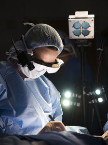
High-tech glasses developed at the School of Medicine help breast surgeon Julie Margenthaler, MD, visualize cancer cells in a patient.

Cancer cells are notoriously difficult to see, even under high-powered magnification. The glasses are designed to make it easier for surgeons to distinguish cancer cells from healthy cells, helping to ensure that no stray tumor cells are left behind during surgery.

“We’re in the early stages of this technology, and more development and testing will be done, but we’re certainly encouraged by the potential benefits to patients,” said breast surgeon Julie Margenthaler, MD, an associate professor of surgery at Washington University, who performed the operation. “Imagine what it would mean if these glasses eliminated the need for follow-up surgery and the associated pain, inconvenience and anxiety.”

The current standard of care requires surgeons to remove the tumor and some neighboring tissue that may or may not include cancer cells. The samples are sent to a pathology lab and viewed under a microscope. If cancer cells are found in neighboring tissue, a second surgery often is recommended to remove additional tissue that also is checked for the presence of cancer.

The glasses could reduce the need for additional surgical procedures and subsequent stress on patients, as well as time and expense.

Margenthaler said about 20-25 percent of breast cancer patients who have lumps removed require a second surgery because current technology doesn’t adequately show the extent of the disease during the first operation. “Our hope is that this new technology will reduce or ideally eliminate the need for a second surgery,” she said.

The technology, developed by a team led by Samuel Achilefu, PhD, professor of radiology and of biomedical engineering at Washington University, incorporates custom video technology, a head-mounted display and a targeted molecular agent that attaches to cancer cells, making them glow when viewed with the glasses.

Samuel Achilefu, PhD

In a study published in the Journal of Biomedical Optics, researchers noted that tumors as small as 1 mm in diameter (the thickness of about 10 sheets of paper) could be detected.

Ryan Fields, MD, a Washington University assistant professor of surgery and Siteman surgeon, plans to wear the glasses later this month when he operates to remove a melanoma from a patient. He said he welcomes the new technology, which theoretically could be used to visualize any type of cancer. “A limitation of surgery is that it’s not always clear to the naked eye the distinction between normal tissue and cancerous tissue,” Fields said. “With the glasses developed by Dr. Achilefu, we can better identify the tissue that must be removed.”

In pilot studies conducted on lab mice, the researchers utilized indocyanine green, a commonly used contrast agent approved by the U.S. Food and Drug Administration. When the agent is injected into the tumor, the cancerous cells glow when viewed with the glasses and a special light.

Achilefu, who also is co-leader of the Oncologic Imaging Program at Siteman Cancer Center and a professor of biochemistry and molecular biophysics, is seeking FDA approval for a different molecular agent he’s helping to develop for use with the glasses. This agent specifically targets and stays longer in cancer cells.

“This technology has great potential for patients and health-care professionals,” Achilefu said. “Our goal is to make sure no cancer is left behind.”

Viktor Gruev, PhD, assistant professor of engineering at WUSTL, and Ron Liang, PhD, of the University of Arizona, assisted with development of the glasses. WUSTL graduate students Suman Mondal, Shengkui Gao and Yang Liu and postdoctoral fellow Nan Zhu also played key roles.


High-tech glasses developed at the School of Medicine help breast surgeon Julie Margenthaler, MD, visualize cancer cells in a patient on Feb. 10. Here is real-time video of the lymph node removal, as seen by Margenthaler as she wore the eyewear. A florescent marker injected into the patient and special lighting made cancer cells glow blue when viewed with the technology. The lighter the shade of blue, the more concentrated the cancer cells are.

Breast cancer vaccine shows promise in small clinical trial

A breast cancer vaccine developed at Washington University School of Medicine in St. Louis is safe in patients with metastatic breast cancer, results of an early clinical trial indicate. Preliminary evidence also suggests that the vaccine primed the patients’ immune systems to attack tumor cells and helped slow the cancer’s progression.

The study appears Dec. 1 in Clinical Cancer Research.

The new vaccine causes the body’s immune system to home in on a protein called mammaglobin-A, found almost exclusively in breast tissue. The protein’s role in healthy tissue is unclear, but breast tumors express it at abnormally high levels, past research has shown.

“Being able to target mammaglobin is exciting because it is expressed broadly in up to 80 percent of breast cancers, but not at meaningful levels in other tissues,” said breast cancer surgeon and senior author William E. Gillanders, MD, professor of surgery. “In theory, this means we could treat a large number of breast cancer patients with potentially fewer side effects.

“It’s also exciting to see this work progress from identifying the importance of mammaglobin-A, to designing a therapeutic agent, manufacturing it and giving it to patients, all by investigators at Washington University,” he added.

The vaccine primes a type of white blood cell, part of the body’s adaptive immune system, to seek out and destroy cells with the mammaglobin-A protein. In the smaller proportion of breast cancer patients whose tumors do not produce mammaglobin-A, this vaccine would not be effective.

In the new study, 14 patients with metastatic breast cancer that expressed mammaglobin-A were vaccinated. The Phase 1 trial was designed mainly to assess the vaccine’s safety. According to the authors, patients experienced few side effects, reporting eight events classified as mild or moderate, including rash, tenderness at the vaccination site and mild flu-like symptoms. No severe or life-threatening side effects occurred.

Although the trial was designed to test vaccine safety, preliminary evidence indicated the vaccine slowed the cancer’s progression, even in patients who tend to have less potent immune systems because of their advanced disease and exposure to chemotherapy.

“Despite the weakened immune systems in these patients, we did observe a biologic response to the vaccine while analyzing immune cells in their blood samples,” said Gillanders, who treats patients at Siteman Cancer Center at Barnes-Jewish Hospital and Washington University. “That’s very encouraging. We also saw preliminary evidence of improved outcome, with modestly longer progression-free survival.”

Of the 14 patients who received the vaccine, about half showed no progression of their cancer one year after receiving the vaccine. In a similar control group of 12 patients who were not vaccinated, about one-fifth showed no cancer progression at the one-year follow-up. Despite the small sample size, this difference is statistically significant.

Based on results of this study, Gillanders and his colleagues are planning a larger clinical trial to test the vaccine in newly diagnosed breast cancer patients, who, in theory, should have more robust immune systems than patients who already have undergone extensive cancer therapy.

“If we give the vaccine to patients at the beginning of treatment, the immune systems should not be compromised like in patients with metastatic disease,” Gillanders said. “We also will be able to do more informative immune monitoring than we did in this preliminary trial. Now that we have good evidence that the vaccine is safe, we think testing it in newly diagnosed patients will give us a better idea of the effectiveness of the therapy.”