Mardis Elected to AACR Board of Directors

Elaine Mardis, PhD, the Robert E. and Louise F. Dunn Distinguished Professor of Medicine, has been elected to the board of directors of the American Association for Cancer Research (AACR).

Mardis, a professor of molecular microbiology and of genetics and co-director of The Elizabeth H. and James S. McDonnell III Genome Institute, will begin her three-year terms at AACR’s annual meeting, April 18-22 in Philadelphia.

For AACR, Mardis also is senior editor of Molecular Cancer Research and a member of the Special Conferences Committee and the Clinical and Translational Cancer Research Steering Committee.

AACR is one of the largest professional organizations dedicated to advancing cancer research. The group publishes peer-reviewed journals and holds conferences that highlight research about the prevention, biology, diagnosis and treatment of cancer.

Kidney Cancer Detected Early With Urine Test

If kidney cancer is diagnosed early — before it spreads — 80 percent of patients survive. However, finding it early has been among the disease’s greatest challenges.

Now, researchers at Washington University School of Medicine in St. Louis have developed a noninvasive method to screen for kidney cancer that involves measuring the presence of proteins in the urine.

The findings are reported March 19 in the journal JAMA Oncology.

The researchers found that the protein biomarkers were more than 95 percent accurate in identifying early-stage kidney cancers. In addition, there were no false positives caused by non-cancerous kidney disease.

“These biomarkers are very sensitive and specific to kidney cancer,” said senior author Evan D. Kharasch, MD, PhD.

Kidney cancer is the seventh most common cancer in men and the 10th most common in women, affecting about 65,000 people each year in the United States. About 14,000 patients die of the disease annually.

Like most cancers, kidney tumors are easier to treat when diagnosed early. But symptoms of the disease, such as blood in the urine and abdominal pain, often don’t develop until later, making early diagnosis difficult.

“The most common way that we find kidney cancer is as an incidental, fortuitous finding when someone has a CT or MRI scan,” said Kharasch, the Russell D. and Mary B. Shelden Professor of Anesthesiology. “It’s not affordable to use such scans as a screening method, so our goal has been to develop a urine test to identify kidney cancer early.”

When kidney cancer isn’t discovered until after it has spread, more than 80 percent of patients die within five years.

With researchers from the Siteman Cancer Center, the Mallinckrodt Institute of Radiology and the Division of Urologic Surgery, Kharasch and principal investigator Jeremiah J. Morrissey, PhD, professor of anesthesiology, analyzed urine samples from 720 patients at Barnes-Jewish Hospital who were about to undergo abdominal CT scans for reasons unrelated to a suspicion of kidney cancer. Results of the scans let the investigators determine whether or not patients had kidney cancer. As a comparison, they also analyzed samples from 80 healthy people and 19 patients previously diagnosed with kidney cancer.

The researchers measured levels of two proteins in the urine — aquaporin-1 (AQP1) and perlipin-2 (PLIN2). None of the healthy people had elevated levels of either protein, but patients with kidney cancer had elevated levels of both proteins.

In addition, three of the 720 patients who had abdominal CT scans also had elevated levels of both proteins. Two of those patients were diagnosed subsequently with kidney cancer, and the third patient died from other causes before a diagnosis could be made.

“Each protein, or biomarker, individually pointed to patients who were likely to have kidney cancer, but the two together were more sensitive and specific than either by itself,” said Morrissey. “When we put the two biomarkers together, we correctly identified the patients with kidney cancer and did not have any false positives.”

Even when patients had other types of non-cancerous kidney disease, levels of the two proteins in the urine were not elevated and did not suggest the presence of cancer.

“Patients with other kinds of cancer or other kidney diseases don’t have elevations in these biomarkers,” Kharasch said. “So in addition to being able to detect kidney cancer early, another advantage of using these biomarkers may be to show who doesn’t have the disease.”

Not all kidney masses found by CT scans turn out to be cancerous, he said. In fact, about 15 percent are not malignant.

“But a CT scan can only tell you whether there is a mass in the kidney, not whether it’s cancer,” Kharasch said. “Currently, the only way to know for sure is to have surgery, and unfortunately, 10 to 15 percent of kidneys removed surgically turn out not to be cancerous.”

Kharasch and Morrissey are working to develop an easy-to-use screening test for kidney cancer, much like mammograms, colonoscopies or other tests designed to identify cancer at early, more treatable stages before patients have symptoms.

“By and large, patients don’t know they have kidney cancer until they get symptoms, such a blood in the urine, a lump or pain in the side or the abdomen, swelling in the ankles or extreme fatigue,” Morrissey said. “And by then, it’s often too late for a cure. Metastatic kidney cancer is extremely difficult to treat, and if the disease is discovered after patients have developed symptoms, they almost always have metastases. So we’re hoping to use the findings to quickly get a test developed that will identify patients at a time when their cancer can be more easily treated.”


Funded by the Barnes-Jewish Hospital Cancer Frontier Fund and The Department of Anesthesiology at Washington University School of Medicine in St. Louis, with additional support from the Bear Cub Fund of Washington University, Barnes-Jewish Hospital Foundation and Washington University Institute of Clinical and Translational Science, with additional funding from the National Cancer Institute (NCI) of the National Institutes of Health (NIH). NIH grant numbers R01CA141521 and UL1 TR000448.

Personalized melanoma vaccines marshal powerful immune response

Personalized melanoma vaccines can be used to marshal a powerful immune response against unique mutations in patients’ tumors, according to early data in a first-in-people clinical trial at Washington University School of Medicine in St. Louis.

The tailor-made vaccines, given to three patients with advanced melanoma, appeared to increase the number and diversity of cancer-fighting T cells responding to the tumors. The finding is a boost to cancer immunotherapy, a treatment strategy that unleashes the immune system to seek out and destroy cancer.

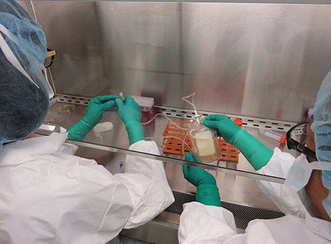
Beatriz Carreno, PhD, (left) and colleague Michelle Becker-Hapak, both of Washington University School of Medicine in St. Louis, deliver a personalized melanoma vaccine into an infusion bag. The vaccine was given to a patient with advanced melanoma via intravenous administration. (Photo: Tom Kitchen)

The research is reported April 2 in Science Express, in a special issue devoted to cancer immunology and immunotherapy.

In a new approach, the cancer vaccines were developed by first sequencing the genomes of patients’ tumors and samples of the patients’ healthy tissues to identify mutated proteins called neoantigens unique to the tumor cells. Then, using computer algorithms and laboratory tests, the researchers were able to predict and test which of those neoantigens would be most likely to provoke a potent immune response and would be useful to include in a vaccine.

The vaccines were given to melanoma patients who had had surgery to remove their tumors but whose cancer cells had spread to the lymph nodes, an indicator the deadly skin cancer is likely to recur. These clinical findings set the stage for a phase I vaccine trial, approved by the Food and Drug Administration as part of an investigational new drug application. The trial will enroll six patients.

Data on the immune response seen in the first three patients is reported in the paper. If additional testing in more patients indicates the vaccines are effective, they may one day be given to patients after surgery to stimulate the immune system to attack lingering cancer cells and prevent a recurrence.

“This proof-of-principle study shows that these custom-designed vaccines can elicit a very strong immune response,” said senior author Gerald Linette, MD, PhD, a Washington University medical oncologist leading the clinical trial at Siteman Cancer Center and Barnes-Jewish Hospital. “The tumor antigens we inserted into the vaccines provoked a broad response among the immune system’s killer T cells responsible for destroying tumors. Our results are preliminary, but we think the vaccines have therapeutic potential based on the breadth and remarkable diversity of the T-cell response.”

It’s too early to say whether the vaccines will be effective in the long term, the researchers cautioned. The study was designed to evaluate safety and immune response; however, none of the patients has experienced adverse side effects.

Earlier attempts at vaccines have focused on targeting normal proteins commonly expressed at high levels in particular cancers. Those same proteins also are found in healthy cells, making it difficult to stimulate a potent immune response.

The new approach investigated by the Washington University team merges cancer genomics with cancer immunotherapy.

“This is about as personalized as vaccines can get,” said co-author Elaine Mardis, PhD, co-director of the McDonnell Genome Institute at Washington University, where the cancer genome sequencing, analysis and neoantigen prediction were performed. “The approach we describe is fundamentally different from conventional mutation discovery, which focuses on identifying mutated genes that drive cancer development. Instead, we’re looking for a unique set of mutated proteins in a patient’s tumor that would be most likely to be recognized by the immune system as foreign.”

Melanomas are notorious for having high numbers of genetic mutations caused by exposure to ultraviolet light. Biopsy samples of melanomas typically carry 500 or more mutated genes. Using prediction algorithms, the researchers narrowed their search for vaccine candidates by identifying neoantigens that not only were expressed in a patient’s tumor but also were likely to be seen by that patient’s immune system as “non-self.”

Biochemical validation of neoantigen peptide expression on the cancer cells’ surfaces was performed in collaboration with William Hildebrand’s group at the University of Oklahoma Health Sciences Center and provided critical assurance that the vaccine would elicit the most effective T cells to combat the melanoma.

“You can think of a neoantigen as a flag on each cancer cell,” said first author Beatriz Carreno, PhD, associate professor of medicine. “Each patient’s melanoma can have hundreds of different flags. As part of validating candidate vaccine neoantigens, we were able to identify the flags on the patients’ cancer cells. Then we created customized vaccines to a select group of flags on each patient’s tumor.”

Carreno and her colleagues selected a set of seven unique neoantigens for each vaccine and used specialized immune cells called dendritic cells, derived from the patients, to carry those neoantigens to the immune system. Dendritic cells play an important role in waking up the immune system, reminding T cells to attack the cancer.

After the vaccine infusions, the patients’ blood was drawn every week for about four months. By analyzing the blood samples, the researchers could see that each patient mounted an immune response to specific neoantigens in their vaccines. The vaccines also stimulated diverse clones of battle-ready T cells against neoantigens, suggesting this approach also could be used to activate a range of T cells and target them to mutations in other cancers with high mutation rates, such as lung cancer, bladder cancer and certain colorectal cancers.

“Our team has developed a new strategy for personalized cancer immunotherapy,” Linette said. “Many researchers have hypothesized that it would be possible to use neoantigens to broadly activate the human immune system, but we didn’t know that for sure until now. We still have much more work to do, but this is an important first step and opens the door to personalized immune-based cancer treatments.”


The research was supported by the Barnes-Jewish Hospital Foundation, Siteman Cancer Frontier Fund, Our Mark on Melanoma Foundation, Come Out Swinging Foundation, Blackout Melanoma Foundation, the National Cancer Institute, grants R21 CA179695 and P30 CA91842, and the National Human Genome Research Institute, grant 5U54HG00307, at the National Institutes of Health (NIH).

Carreno BM, Magrini V, Becker-Hapak M, Kaabinejadian S, Hundal J, Petti AA, Ly A, Lie W-R, Hildebrand WH, Mardis ER and Linette GP. A dendritic cell vaccine increases the breadth and diversity of melanoma neoantigen- specific T cell. Science Express, April 2, 2015.

Innovative Light Therapy Reaches Deep Tumors

Light long has been used to treat cancer. But phototherapy is only effective where light easily can reach, limiting its use to cancers of the skin and in areas accessible with an endoscope, such as the gastrointestinal tract.

Using a mouse model of cancer, researchers at Washington University School of Medicine in St. Louis and the Siteman Cancer Center have devised a way to apply light-based therapy to deep tissues never before accessible. Instead of shining an outside light, they delivered light directly to tumor cells, along with a photosensitive source of free radicals that can be activated by the light to destroy cancer. And they accomplished this using materials already approved for use in cancer patients.

The study appears March 9 in the journal Nature Nanotechnology.

“Phototherapy works very well and has few side effects, but it can’t be used for deeply embedded or metastatic tumors,” said senior author Samuel Achilefu, PhD, professor of radiology and of biomedical engineering at Washington University. “In general, shining a light on photosensitive materials generates free radicals that are very toxic and induce cell death. But the technique has only worked well when light and oxygen can get there. The need for oxygen and the shallow penetration of light in tissue have limited advances in this area for decades.”

The light source the researchers harnessed relies on a phenomenon called Cerenkov radiation, identified in the 1930s by Pavel Cerenkov, who later won the Nobel Prize in Physics for the discovery. Cerenkov radiation is responsible for the characteristic blue glow of underwater nuclear reactors. It also is produced during positron emission tomography (PET) scans that doctors use to diagnose cancer.

Achilefu and first author Nalinikanth Kotagiri, MD, PhD, a postdoctoral researcher, focused on a widely used imaging strategy called FDG-PET. With this technique, patients undergo a PET scan after receiving an intravenous dose of radiolabeled sugar molecules called fluorodeoxyglucose (FDG). Many tumors take up the sugar to support their rapid growth, and the attached radioactive fluorine makes those tumors light up on a PET scan, no matter where they are in the body.

The researchers hypothesized that the radioactive fluorine also would produce enough Cerenkov radiation to activate a photosensitizing agent if it could also be delivered to the same location.

In this way, FDG could serve two purposes, continuing its role as an imaging agent and adding the new job of providing light for phototherapy, according to Kotagiri.

“FDG is one of the most widely used imaging agents in the world,” Achilefu said. “That’s the beauty of this treatment paradigm. It’s used in hospitals today to find primary and metastatic cancer. So with FDG as our light source, we needed to find a material that becomes toxic when exposed to the light it produces.”

After looking at a number of options, the researchers focused on nanoparticles made of titanium dioxide, a mineral with wide applications in medicine and industry including in hip implants, sunscreen, toothpaste and food additives. When exposed to light, titanium dioxide produces free radicals without requiring oxygen for the reaction. To see if they could increase the potency of the nanoparticles, the investigators also added a drug called titanocene to the nanomaterial’s surface.

“Titanocene has been approved for investigational use in people,” Achilefu said. “It went all the way to Phase 2 clinical trials as a chemotherapy agent. It was found to be safe, but it didn’t work that well compared with a placebo. Still, it’s also known to interact with low-intensity light and break into free radicals. We decided to see if we could teach it to do its job differently — to act as a phototherapeutic drug instead of a chemotherapeutic drug.”

The titanium dioxide nanoparticle is shown here (purple) carrying the iron-binding protein transferrin (blue and green) and the light-sensitive cancer drug titanocene (red). (Credit: Kotagiri)

To help the nanoparticles home in on tumors in mice, the researchers also coated the particles with a protein called transferrin that binds to iron in the blood. Like sugar, many tumors rely on iron to grow. Achilefu pointed out that this iron-binding protein is simply one example of a way to target the photosensitive materials to cancer cells.

The researchers tested different formulations of the nanoparticles and cancer drug combined with the FDG light source in mice with human lung tumors and fibrosarcoma, a tumor of the connective tissue. Comparing these mice with untreated mice, they tested the following combinations: FDG plus tumor-seeking nanoparticles alone (no cancer drug), FDG plus tumor-seeking cancer drug alone (no nanoparticles), and FDG plus tumor-seeking nanoparticles carrying the cancer drug.

When injected into the bloodstream with FDG, the tumor-seeking nanoparticles that carried the cancer drug had the most significant effect. Fifteen days after treatment, tumors in treated mice were eight times smaller than those in untreated mice.

Mice that received FDG plus tumor-seeking nanoparticles alone survived about 30 days compared to an average of 15 days for untreated mice. They also found about the same 30-day survival for mice that received FDG plus just the tumor-seeking cancer drug — without the nanoparticles. Survival increased to 50 days for mice receiving all three components: FDG plus the tumor-seeking nanoparticles carrying the cancer drug.

“Exposed to the light source, the titanium dioxide nanoparticles alone can kill cancer,” Achilefu said. “But adding the drug appears to enhance the therapeutic outcome. The two together produce different kinds of free radicals that overwhelm tumor cells. Our formulation also uses doses of the drug that are much lower than would be administered for chemotherapy.”

Kotagiri added that toxic side effects should be minimal. Both the light and the photosensitive material are targeted to the tumor, and the material is not toxic unless activated by the light source, which should occur only at the tumor site.

Achilefu and Kotagiri are planning a small clinical trial in people to evaluate the readily available components of this strategy, beginning with FDG combined with the investigational cancer drug.


This work was supported in part by the National Institutes of Health (NIH), grant numbers R01 CA171651, P50 CA094056, R01 EB008111 and SIG S10 RR031626; and by the National Science Foundation (NSF), grant number CCF 0963742.

Kotagiri N, Sudlow GP, Akers WJ, Achilefu S. Breaking the depth dependency of phototherapy with Cerenkov radiation and low radiance responsive nanophotosensitizers. Nature Nanotechnology. March 9, 2015.

Preventing early-onset colorectal cancers aim of $25 million award

School of Medicine leads international team funded by Cancer Grand Challenges

Colorectal cancer rates in adults under age 50 have been increasing in the U.S. and globally. The risk of developing such cancers has been rising with each generation since the 1950s, but scientists have not been able to explain why. Understanding the underlying causes will help design prevention strategies for future generations.

Yin Cao, ScD, an associate professor of surgery and of medicine in the Public Health Sciences Division at Washington University School of Medicine in St. Louis, is leading an international team seeking to understand what is driving the increase in young-onset colorectal cancers globally. Her team will do so with an award of up to $25 million over five years from Cancer Grand Challenges, a global research funding initiative co-founded by Cancer Research UK and the National Cancer Institute (NCI), part of the National Institutes of Health (NIH). According to the organization, the mission of Cancer Grand Challenges is to unite the world’s brightest minds against cancer’s toughest challenges.

The team is funded by Cancer Research UK, the National Cancer Institute, the Bowelbabe Fund for Cancer Research UK, and Institut National Du Cancer in France, through Cancer Grand Challenges.

“The alarming rise of colorectal cancer in young people around the world demands immediate action,” said Cao, also a research member of Siteman Cancer Center, based at Barnes-Jewish Hospital and Washington University School of Medicine. “We used to think colorectal cancer was a disease of the older population, but in the past two decades, the median age of diagnosis for this cancer in the U.S. dropped from 72 to 66 years. Our team aims to unravel and ultimately reverse the intricate network of causal factors throughout the life course that contribute to early-onset colorectal cancer.”

The award will provide funding for Cao’s research team — named Pathways, Risk Factors and Molecules to Prevent Early-onset Colorectal Tumors (PROSPECT) — to develop disruptive and transdisciplinary approaches to address this challenge. Among 176 international teams that applied, Cao’s team is one of five to receive funding this year through Cancer Grand Challenges. The other funded teams are focused on cancer inequities, solid tumors in children, and T cell receptors.

Cao co-leads the team with Andrew Chan, MD, at Massachusetts General Hospital (MGH). The research team also involves nine interdisciplinary co-investigators across the globe, including Gary Patti, PhD, the Michael and Tana Powell Professor of Chemistry at Washington University. Patti, a pioneer of mass spectrometry-based technologies, will lead state-of-the-art molecular and metabolic analyses to identify possible new risk factors.

Cao’s lab has led the field of early-onset colorectal cancer research by uncovering risk factors throughout the life course that may contribute to the cancer’s rising incidence, such as obesity; diabetes; metabolic comorbid conditions such as high blood pressure and high cholesterol; prolonged sitting; poor diet; sugar-sweetened beverages; alcohol drinking patterns; and birth via cesarean delivery. Cao’s team also leads work in early detection and understanding molecular pathways of this cancer.

Building upon this past research, Cao will lead the first consortium dedicated to early-onset colorectal cancer risk factor discovery. The PROSPECT Global Consortium will have support from more than 15 international cohorts and electronic health record-based biobanks from high-, middle- and low-income countries. Her team will examine known risk factors and seek to find new ones. The query will encompass environmental exposures throughout the life course, including lifestyle, genetic, environmental and social factors. The team then will characterize possible causal factors and the cumulative impact of exposures over the life course on colorectal cancer initiation and progression, using animal models.

Cao also will also collaborate with clinicians at Siteman Cancer Center’s Young Onset Colorectal Cancer Program to collect biospecimens along the continuum of early-onset colorectal cancer development. The researchers will analyze these specimens to characterize the cumulative effects of risk factors for early-onset colorectal cancer on the biochemical and molecular states of tissues.

“An understanding of what causes early-onset cancers is important for the development of interventions based on an individual’s risk so that we can prevent disease and death,” Cao said. “Our goal is to maximally integrate human and mechanistic evidence to develop precision prevention strategies.”

In addition to identifying risk factors and evaluating causal factors, the team also will translate these findings to develop and test prevention strategies, including precision prevention trials and community risk-assessment trials.

“By pushing the boundaries of our individual fields, we hope to move quickly to identify opportunities for preventive interventions that can benefit younger populations,” Cao said. “It’s a joint adventure shared by scientists, providers, patients and the public under a unified goal: shaping a hopeful and healthier future for our younger generations.”

About Washington University School of Medicine

WashU Medicine is a global leader in academic medicine, including biomedical research, patient care and educational programs with 2,900 faculty. Its National Institutes of Health (NIH) research funding portfolio is the second largest among U.S. medical schools and has grown 56% in the last seven years. Together with institutional investment, WashU Medicine commits well over $1 billion annually to basic and clinical research innovation and training. Its faculty practice is consistently within the top five in the country, with more than 1,900 faculty physicians practicing at 130 locations and who are also the medical staffs of Barnes-Jewish and St. Louis Children’s hospitals of BJC HealthCare. WashU Medicine has a storied history in MD/PhD training, recently dedicated $100 million to scholarships and curriculum renewal for its medical students, and is home to top-notch training programs in every medical subspecialty as well as physical therapy, occupational therapy, and audiology and communications sciences.

Author: Marley Wiemers, Contributing Science Writer

Media Contact: James Goodwin, Associate Director of Strategic Communication, Siteman Cancer Center,

314-680-8251 | [email protected]

$2.1 million in cancer research grants awarded by Siteman Investment Program

Research on colon cancerbreast cancer and acute myeloid leukemia are among the seven projects that will benefit from $2.1 million in new grants announced by Siteman Cancer Center through its Siteman Investment Program. The goal of the grants is to support and accelerate the pace of innovation in cancer research.

The money awarded comes from a variety of sources: Pedal the Cause annual bike challenge and Illumination gala, through the Cancer Frontier Fund at the Foundation for Barnes-Jewish Hospital; Fashion Footwear Association of New York; National Cancer Institute; and Barnard Trust.

The research projects are described below.

Title: Pilot Implementation and Feasibility Assessment: Multilevel Intervention to Reduce Rural Colon Cancer Disparities

Principal investigator: Aimee James, PhD, an associate professor of surgery at Washington University School of Medicine and a research member of Siteman Cancer Center

Goal: To develop a multi-level intervention to improve colon cancer screening and follow-up in primary care clinics in rural Southern Illinois

Summary: Rural areas in the Midwest face higher rates of colon cancer mortality. Screening can help reduce the burden, but screening rates are relatively low in rural areas. Further, many people who get screened do not receive appropriate diagnostic follow-up, especially if they were screened with fecal testing. Better and more consistent implementation of strategies that we know can be effective can help underserved communities increase screening and improve follow-up of abnormal cancer screening tests. Interventions at multiple levels are critical to success, as intervening on patients or primary care providers alone is unlikely to have substantial impact. We have partnered with Southern Illinois Healthcare to develop a multi-level intervention using evidence-based strategies to improve screening and follow-up in primary care clinics in rural Southern Illinois.

Title: Retinoid Therapy in Acute Myeloid Leukemia

Principal investigator: John Welch, MD, PhD, an associate professor of medicine at Washington University School of Medicine and a research member of Siteman Cancer Center

Goal: To determine if two existing acute myeloid leukemia (AML) drugs are more effective when administered in combination with each other using mouse and human models

Summary: This project focuses on completing the required pre-clinical studies needed to translate a benchtop observation into a clinical trial in acute myeloid leukemia (AML). We found that two old drugs, all-trans retinoic acid and bexarotene, exhibit strong synergy when added together in a mouse model of AML, whereas they have very modest activity by themselves. Both drugs are FDA approved, are orally available and have well-characterized and tolerable side effects. In this proposal, we will determine whether this combination is highly active in other mouse and human models of AML, or if these drugs are only active in this one mouse model of AML (MLL-AF9). In addition, we will work with medicinal chemists at Washington University to make multiple subtle changes in the chemical structure of bexarotene, and determine whether any of these changes augment the synergistic interaction and could improve on bexarotene as anti-leukemic agents. These results will form the justification for future clinical trials in AML.

Title: Oral Microbiome, Virome and Barrett’s Esophagus

Principal investigator: Yin Cao, ScD, an assistant professor of surgery at Washington University School of Medicine

Goal: To better understand the relationship between bacteria in one’s oral cavity and a person’s future risk of developing Barrett’s esophagus and esophageal adenocarcinoma

Summary: In the U.S., esophageal adenocarcinoma (EAC) has had a nearly seven-fold increase in incidence over the last four decades. However, EAC is highly lethal compared to other cancers, in that 80 percent of EAC patients succumb to the disease within 5 years. The prevention and early detection of EAC and Barrett’s esophagus (BE), the established premalignant lesion of EAC, is a high clinical priority and research challenge. Bacteria and virus may be important to this disease process but studies are limited. Project 1 will, for the first time, examine bacteria in the oral cavity and how that relates to future risk of BE in two large studies that have followed an initially healthy population for more than 30 years. Project 2 will look into whether viruses in the saliva samples collected from the above studies could be detected, as well as from esophageal samples collected from BE patients in a large hospital based cohort. The two projects will provide novel knowledge on BE/EAC development.

Title: Radio-sensitization by Free Fatty Acid Supplementation in Cervical Cancer

Principal investigator: Julie Schwarz, MD, PhD, an associate professor of radiation oncology at Washington University School of Medicine and a research member of Siteman Cancer Center

Goal: To explore why obese patients treated with radiation therapy for cervical cancer have better response rates than patients who are not obese, with the ultimate goal of identifying a dietary supplement or drug that can be given to patients to promote better treatment responses

Summary: Many patients struggle with obesity, and studies have shown that obese cancer patients treated with surgery and chemotherapy have poor outcomes. Surprisingly, we have found that obese patients treated with radiation therapy for cervical cancer are cured more often than patients who are not obese. Obese patients have increased body fat and circulating levels of fats. In the laboratory, we have shown that free fatty acids enhance the effects of radiation. After radiation treatment, cancer cells actively take up fatty acids, and this results in changes in cell signaling that promote tumor cell death. In this grant, we will perform additional experiments in cells and animal models to understand how free fatty acids and obesity are promoting the response to radiation therapy. Our ultimate goal is to identify a dietary supplement or drug that we can give to patients that will promote treatment sensitivity.

Title: Intravenous Lidocaine for Preventing Oxaliplatin-induced Peripheral Neuropathy (OIPN)

Principal investigator: Simon Haroutounian, PhD, an assistant professor of anesthesiology at Washington University School of Medicine

Goal: To determine if lidocaine can reduce the occurrence and severity of oxaliplatin-induced peripheral neuropathy, a painful common side effect of chemotherapy

Summary: Colorectal cancer is the second leading cause of cancer-related death in the U.S. Oxaliplatin is a key chemotherapy agent demonstrating improved survival in colorectal cancer, but causes injury to sensory nerves in 72 percent of patients receiving treatment. This nerve damage, called peripheral neuropathy (PN), can cause substantial pain, numbness and sensory changes such as extreme sensitivity to cold. PN leads to dose reduction or early discontinuation of oxaliplatin in 50 to 70 percent of patients, reducing patient survival by 5 to 14 months. In approximately 20 percent of patients, oxaliplatin-induced PN persists for months or years after chemotherapy. The study aims to determine whether intravenous lidocaine can reduce the incidence and the severity of oxaliplatin-induced PN. In a prospective, randomized study, patients with colorectal cancer will receive lidocaine or placebo with their standard-of-care oxaliplatin-based chemotherapy. The key outcomes, compared between the groups, include: 1) symptoms and signs of peripheral neuropathy, 2) cumulative dose of oxaliplatin and 3) adverse effects.

Title: Genomic Classification and Targeted Immunotherapy of Disseminated Tumor Cells in Breast Cancer Patients

Principal investigators: Siteman Cancer Center research members Rebecca Aft, MD, PhD, a professor of surgery; Mark Watson, MD, PhD, an associate professor of pathology and immunology; and Leonel Hernandez-Aya, MD, an assistant professor of medicine, all of Washington University School of Medicine

Goal: To better understand how to develop new therapies to eliminate disseminated tumor cells in triple negative breast cancer patients, to prevent the cancer from metastasizing

Summary: Treatment of triple negative breast cancer is challenging since there are a limited number of new and highly effective therapies. This subset of breast cancer patients has a high risk of disease recurrence and death when traditional chemotherapy is used. Small deposits of disseminated tumor cells released from the breast tumor, detected in the bone marrow of some triple negative breast cancer patients before and after chemotherapy treatment, identify patients with a particularly high risk of developing metastatic disease. It is thought that metastatic disease develops in these patients because these tumor cells themselves are resistant to therapy and because other environmental factors selectively support their growth and survival. We have developed a very sensitive 8-gene molecular biomarker panel that can identify triple negative breast cancer patients who have disseminated tumor cells in their bone marrow and who have a high risk of cancer recurrence. In this project, we will isolate and further characterize the genetics of individual disseminated tumor cells from triple negative breast cancer patients, to better understand how to develop new therapies to eliminate them and prevent metastatic disease. We will also examine whether and how a patient’s own immune cells contribute to the survival of disseminated tumor cells in the bone marrow. Importantly, we will conduct a pilot clinical trial to determine whether altering the function of immune cells in a patient’s bone marrow can help eliminate these tumor cells and prevent future metastatic tumor recurrence. Overall, these studies are geared to discover novel therapeutic strategies to eliminate disseminated tumor cells and potentially improve long-term survival in patients with triple negative breast cancer.

Title: Advancing Therapies for Incurable Lymphomas via Translational Team Science

Principal investigator: Siteman Cancer Center research members Todd Fehniger, MD, PhD, an associate professor of medicine and Brad Kahl, MD, a professor of medicine, both of Washington University School of Medicine

Goal: To develop and evaluate new treatments for patients with incurable lymphomas

Summary: The long-term goals are to advance novel basic findings in lymphoma into the clinic via development of a comprehensive and innovate translational research program. Specific projects seek to:

  1. Investigate new immunotherapy treatment combinations in early phase clinical trials, to boost indolent lymphoma patients’ natural killer, or NK, cells, and to further enhance NK cell targeting of lymphoma cells via modification with specialized chimeric antigen receptors (CAR);
  2. Understand how changes in lymphoma cells’ DNA result in follicular lymphoma, and translate these genetic findings into new ways to help with lymphoma patient prognosis and develop new personalized lymphoma-specific vaccine therapies;
  3. Develop next-generation “universal” CAR T cells to reduce barriers to lymphoma patients receiving CAR-modified T cellular therapy, and explore new strategies to enhance CAR-T cell persistence;
  4. Determine how changes in the structure of DNA (epigenetic) impact response to treatment and progression in very hard-to-treat T cell lymphomas; and
  5. Understand the role of key activated receptor pathways (growth signals) for aggressive T-cell lymphoma/leukemia, and advance a new combination clinical trial of chemotherapy combined with a growth pathway inhibitor.

These projects will include or lead to new clinical trials for lymphoma patients, with the ultimate goal of curing lymphoma.