


The Siteman Cancer Center is committed to early detection of breast cancer through mammography and other screening methods.
Trust starts with a dedicated team of WashU Medicine radiologists affiliated with the internationally recognized Mallinckrodt Institute of Radiology.
Expertise in Breast Cancer Imaging
Where you get your mammogram makes a difference. At Siteman Cancer Center, every mammogram is read by an imaging expert with the skill, knowledge and experience only a national leader can deliver. At Siteman, our radiologists are part of a team of WashU Medicine physicians that includes sub-specialized breast health experts.
All radiology technologists on staff have advanced certification in breast mammography and have undergone rigorous training in breast anatomy and physiology, positioning and compression, as well as imaging of patients with breast implants.
Their expertise and experience means clear images the first time and every time. At Siteman, we provide all digital breast imaging services including:
- Screening mammograms, including 3D mammography (tomosynthesis)
- Diagnostic mammograms
- Breast ultrasound
- Breast Magnetic Resonance Imaging (MRI)
- Minimally invasive breast biopsy
Make an Appointment
Use the buttons below to schedule your mammogram online with the BJC Online Self-Scheduler, or call to request an appointment. Our Siteman care coordinators – oncology-trained nurses who help with scheduling, finding the right specialist, and guidance – can help you through the process.
Locations
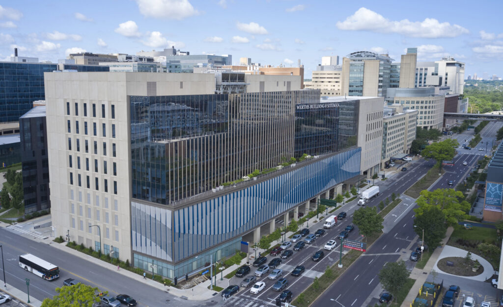


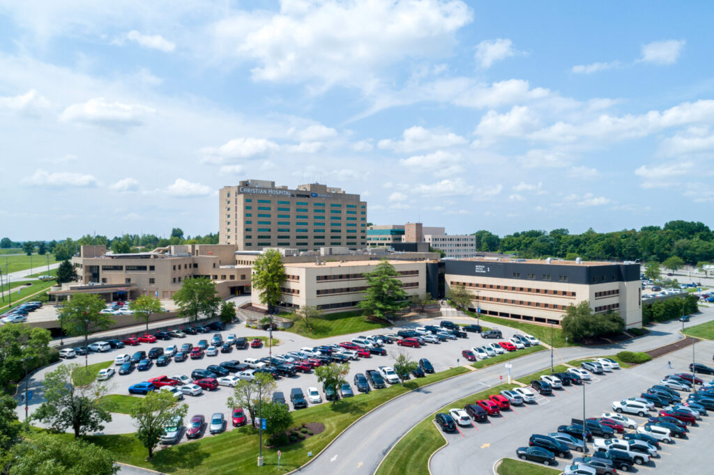
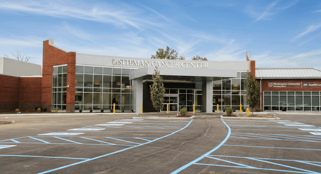
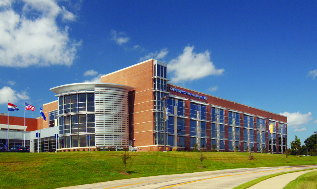




Services for Underinsured and Uninsured Women
No-cost mammograms are available for underinsured and uninsured women who qualify through the Breast Health Outreach Program at the Joanne Knight Breast Health Center. The outreach program is the largest and most comprehensive of its kind in the state of Missouri. Please call 314-454-8466 for more information.
Calculate your breast cancer risk
Your Disease Risk® is a tool developed by our experts that can help you determine your risk of breast cancer and 11 other cancers.
Breast Health FAQs
One of the best tools we have in the fight against breast cancer is screening mammography, which has helped reduce breast cancer mortality in the U.S. by nearly 40 percent since 1990. Mammography saves lives – read on to find out what you need to know.
General mammography
A traditional screening mammogram is an imaging test that looks for breast cancer in women who are considered at average risk for the disease and who have no signs or symptoms. Screening mammograms usually involve two or more low-dose x-ray pictures of each breast.
A 3D mammogram (also known as breast tomosynthesis) is a screening exam that involves multiple x-ray images of the breast taken from different angles. Then, a computer uses those images to create a 3D representation of the breast. The benefit of tomosynthesis is that it separates overlapping tissues, making it easier to see abnormalities. Almost all of our screening mammograms are performed as 3D mammograms these days.
While a screening mammogram looks for breast cancer in women with no symptoms, a diagnostic mammogram is meant to evaluate women who have clinical symptoms (like a lump) or to look more closely at something that is seen on a screening mammogram. Diagnostic mammograms involve different x-ray pictures than the standard images taken during a screening mammogram. A diagnostic evaluation may also include an ultrasound. You will get the results of your diagnostic mammogram before you leave the facility.
Screening mammograms are able to catch breast cancer early, when it’s easier to treat. Regular mammograms can lower the risk of dying from breast cancer. According to the Centers for Disease Control and Prevention, a mammogram is the best way to find breast cancer in most women of screening age.
If you’ve already noticed any changes in your breast, you may need a diagnostic mammogram to investigate the cause. Diagnostic mammograms are also used to evaluate abnormal results from a screening mammogram.
Breast ultrasound isn’t typically used as a screening tool for breast cancer. Instead, an ultrasound may be performed if the results of a screening mammogram were abnormal. A screening mammogram is used to look for breast cancer when there are no symptoms. A breast ultrasound, on the other hand, is used as a diagnostic tool to evaluate a problem found during a physical exam or a screening mammogram.
Breast cancer risk and screening
Even if you don’t have a family history of breast cancer, the National Comprehensive Cancer Network (NCCN) still recommends that women at average risk for breast cancer begin screening mammograms at age 40. In fact, most women who get breast cancer don’t have a family history of the disease, so it’s important to get screened annually.
Siteman recommends that women begin annual screening mammograms at age 40. We follow NCCN guidelines for our patients.
Annual screening at age 40 is also recommended for:
- Transgender women (male-to-female), nonbinary and genderqueer patients who have used gender-affirming hormones for longer than five years
- Transgender men (female-to-male), nonbinary and genderqueer patients whose sex assigned at birth is female who haven’t had top surgery
Breasts need to be compressed during mammograms to optimize the image quality and makes it easier for radiologists to see through breast tissue. It also minimizes the amount of radiation exposure to the breast.
Your mammogram will be performed by a technologist called a mammographer. They are specially trained to perform mammograms, ensuring that high-quality images are taken while minimizing the amount of radiation exposure.
At Siteman, all radiology technologists have advanced certification in breast mammography and have undergone rigorous training in breast anatomy and physiology, positioning and compression.
Mammography results
Your images will be interpreted by a board-certified radiologist with expertise in breast imaging. Radiologists are physicians who are specially trained to interpret images to diagnose diseases and injuries.
WashU Medicine radiologists are affiliated with the internationally recognized Mallinckrodt Institute of Radiology.
Breast density varies greatly from person to person. Some breasts have almost all fatty tissue, while others have extremely dense tissue with very little fat. The denser the breast, the harder it can be to see masses or other findings on a mammogram. This is because both dense breast tissue and masses appear as white areas on images.
If you have dense breasts, you still need regular mammograms. While dense breast tissue can make it difficult to see cancer, most breast cancers can still be seen on a mammogram. And, according to the NCCN, women who have dense breasts are at higher risk for breast cancer, so it’s especially important to keep up with regular screening.
After the radiologist interprets your images, they will send your results to you. They will also send your results to your primary care or referring physician, who will go over your results with you.
While getting called back after a screening mammogram can be scary, it’s actually quite common. Most women who get called back don’t have cancer. In fact, fewer than one in ten women who are called back for follow-up testing end up having cancer.
Follow-up tests that may be used to rule out cancer include a diagnostic mammogram, a breast ultrasound or an MRI. Based on the results of your follow-up test, you may need a biopsy to confirm a diagnosis.
Mammography safety concerns
Yes! If you have breast implants, you should still get screened regularly according to breast cancer screening recommendations. It’s important to tell your technologist if you have breast implants before your exam begins. Both silicone and saline implants can make it difficult for the radiologist to see breast tissue. For this reason, women with implants have four extra images taken in addition to the four standard mammogram images.
Yes, mammography can be performed during pregnancy. The risk of radiation to the fetus is extremely low. A lead shield is placed over the belly to keep radiation from reaching the womb. If you’re pregnant, or think you could be, make sure to tell your technologist before your mammogram.
If you have concerns about getting your mammogram, you can speak with your referring provider. However, experts agree that mammography during pregnancy is safe.
Yes, you can get a mammogram while you’re breastfeeding. There is no risk to your baby/child. We do recommend that you nurse or pump immediately before your mammogram to allow for the best images. If you’re breastfeeding, make sure to tell your technologist before your mammogram.
The dose of radiation used during a mammogram is extremely low – lower than any other x-ray or CT scan. Mammography is very safe and special care is taken to make sure that you’ll be exposed to the smallest amount possible. The benefits of mammography outweigh any possible harm from this minimal radiation exposure.
Insurance
Almost all health insurance plans cover the cost of screening mammograms and other cancer screenings. Be sure to call your insurance carrier before getting screened to confirm that your mammogram will be covered.
Diagnostic mammograms and ultrasounds typically are not fully covered as routine preventive health, and are subject to copayment and deductibles, per your specific plan.
If you’re uninsured or underinsured and are concerned about the cost, BJC HealthCare offers financial assistance.
Learn more about insurance and cancer screenings.